 Movie
Movie Controller


+ Open data

- Basic information
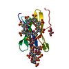
| Entry | Database: PDB / ID: 9hbg | |||||||||
|---|---|---|---|---|---|---|---|---|---|---|
| Title | The PK-RSL - phosphonato-calix[6]arene cocrystal structure | |||||||||
 Components Components | Fucose-binding lectin protein | |||||||||
 Keywords Keywords | SUGAR BINDING PROTEIN / Beta-propeller / lectin / trimer | |||||||||
| Function / homology | Fucose-specific lectin / Fungal fucose-specific lectin / carbohydrate binding / metal ion binding / phosphonato-calix[6]arene / beta-D-fructopyranose / Fucose-binding lectin protein Function and homology information Function and homology information | |||||||||
| Biological species |  Ralstonia solanacearum (bacteria) Ralstonia solanacearum (bacteria) | |||||||||
| Method |  X-RAY DIFFRACTION / SYNCHROTRON / MOLECULAR REPLACEMENT / Resolution: 1.28 Å X-RAY DIFFRACTION / SYNCHROTRON / MOLECULAR REPLACEMENT / Resolution: 1.28 Å | |||||||||
 Authors Authors | Mockler, N.M. / Crowley, P.B. | |||||||||
| Funding support |  Ireland, 2items Ireland, 2items
| |||||||||
 Citation Citation | Journal: Chemistry / Year: 2025 Title: Making and Breaking Supramolecular Synthons for Modular Protein Frameworks. Authors: Mockler, N.M. / Raston, C.L. / Crowley, P.B. | |||||||||
| History |
|
- Structure visualization
| Structure viewer | Molecule: MolmilJmol/JSmol |
|---|
- Downloads & links
-Download
| PDBx/mmCIF format | 9hbg.cif.gz | 44.9 KB | Display | PDBx/mmCIF format |
|---|---|---|---|---|
| PDB format | pdb9hbg.ent.gz | 24.7 KB | Display | PDB format |
| PDBx/mmJSON format | 9hbg.json.gz | Tree view | PDBx/mmJSON format | |
| Others |  Other downloads Other downloads |
-Validation report
| Arichive directory | https://data.pdbj.org/pub/pdb/validation_reports/hb/9hbgftp://data.pdbj.org/pub/pdb/validation_reports/hb/9hbg | HTTPS FTP |
|---|
-Related structure data


-Links
 PDBj
PDBj- Assembly
| Deposited unit | 
| ||||||||||||
|---|---|---|---|---|---|---|---|---|---|---|---|---|---|
| 1 |
| ||||||||||||
| Unit cell |
| ||||||||||||
| Components on special symmetry positions |
|
-Components
| #1: Protein | Mass: 9872.778 Da / Num. of mol.: 1 / Mutation: Pro0, S1K Source method: isolated from a genetically manipulated source Details: Ralstonia solanacearum lectin with a Pro-Lys N-terminus Source: (gene. exp.) Ralstonia solanacearum (bacteria) / Gene: E7Z57_08365, HF909_06975, LBW55_09125, RUN39_v1_50103 / Production host: | ||||||||
|---|---|---|---|---|---|---|---|---|---|
| #2: Sugar |   Type: D-saccharide, beta linking / Mass: 180.156 Da / Num. of mol.: 2 / Source method: obtained synthetically / Formula: C6H12O6 / Feature type: SUBJECT OF INVESTIGATION Type: D-saccharide, beta linking / Mass: 180.156 Da / Num. of mol.: 2 / Source method: obtained synthetically / Formula: C6H12O6 / Feature type: SUBJECT OF INVESTIGATION#3: Chemical | ChemComp-7AZ / | 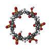  Mass: 1116.611 Da / Num. of mol.: 1 / Source method: obtained synthetically / Formula: C42H42O24P6 / Feature type: SUBJECT OF INVESTIGATION Mass: 1116.611 Da / Num. of mol.: 1 / Source method: obtained synthetically / Formula: C42H42O24P6 / Feature type: SUBJECT OF INVESTIGATION#4: Water | ChemComp-HOH / |  Mass: 18.015 Da / Num. of mol.: 120 / Source method: isolated from a natural source / Formula: H2O Mass: 18.015 Da / Num. of mol.: 120 / Source method: isolated from a natural source / Formula: H2OHas ligand of interest | Y | Has protein modification | N | |
-Experimental details
-Experiment
| Experiment | Method: X-RAY DIFFRACTION / Number of used crystals: 1 |
|---|
- Sample preparation
| Crystal | Density Matthews: 2.62 Å3/Da / Density % sol: 53 % |
|---|---|
| Crystal grow | Temperature: 293 K / Method: vapor diffusion, hanging drop / pH: 4.5 Details: 1.1 M Di-ammonium hydrogen phosphate, 0.1 M sodium acetate pH 4.5 |
-Data collection
| Diffraction | Mean temperature: 100 K / Serial crystal experiment: N |
|---|---|
| Diffraction source | Source: SYNCHROTRON / Site: SOLEIL  / Beamline: PROXIMA 2 / Wavelength: 0.98 Å / Beamline: PROXIMA 2 / Wavelength: 0.98 Å |
| Detector | Type: DECTRIS EIGER X 9M / Detector: PIXEL / Date: Nov 16, 2021 |
| Radiation | Monochromator: M / Protocol: SINGLE WAVELENGTH / Monochromatic (M) / Laue (L): M / Scattering type: x-ray |
| Radiation wavelength | Wavelength: 0.98 Å / Relative weight: 1 |
| Reflection | Resolution: 1.28→31.25 Å / Num. obs: 29915 / % possible obs: 100 % / Redundancy: 39 % / Biso Wilson estimate: 20 Å2 / CC1/2: 1 / Rmerge(I) obs: 0.048 / Net I/σ(I): 37.6 |
| Reflection shell | Resolution: 1.28→1.298 Å / Rmerge(I) obs: 1.556 / Num. unique obs: 1478 / CC1/2: 0.827 |
- Processing
| Software |
| ||||||||||||||||||||||||||||||||||||||||||||||||||||||||||||||||||||||||||||||||||||
|---|---|---|---|---|---|---|---|---|---|---|---|---|---|---|---|---|---|---|---|---|---|---|---|---|---|---|---|---|---|---|---|---|---|---|---|---|---|---|---|---|---|---|---|---|---|---|---|---|---|---|---|---|---|---|---|---|---|---|---|---|---|---|---|---|---|---|---|---|---|---|---|---|---|---|---|---|---|---|---|---|---|---|---|---|---|
| Refinement | Method to determine structure: MOLECULAR REPLACEMENT / Resolution: 1.28→31.25 Å / SU ML: 0.1699 / Cross valid method: FREE R-VALUE / σ(F): 1.37 / Phase error: 30.6373 Stereochemistry target values: GeoStd + Monomer Library + CDL v1.2
| ||||||||||||||||||||||||||||||||||||||||||||||||||||||||||||||||||||||||||||||||||||
| Solvent computation | Shrinkage radii: 0.9 Å / VDW probe radii: 1.1 Å / Solvent model: FLAT BULK SOLVENT MODEL | ||||||||||||||||||||||||||||||||||||||||||||||||||||||||||||||||||||||||||||||||||||
| Displacement parameters | Biso mean: 27.61 Å2 | ||||||||||||||||||||||||||||||||||||||||||||||||||||||||||||||||||||||||||||||||||||
| Refinement step | Cycle: LAST / Resolution: 1.28→31.25 Å
| ||||||||||||||||||||||||||||||||||||||||||||||||||||||||||||||||||||||||||||||||||||
| Refine LS restraints |
| ||||||||||||||||||||||||||||||||||||||||||||||||||||||||||||||||||||||||||||||||||||
| LS refinement shell |
|
