 Movie
Movie Controller

[English] 日本語
 Yorodumi
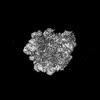
- PDB-9ha7: Pooled 50S subunit C-CP_(L22)-~H61 precursor states supplemented ... -
+ Open data

- Basic information
| Entry | Database: PDB / ID: 9ha7 | |||||||||||||||
|---|---|---|---|---|---|---|---|---|---|---|---|---|---|---|---|---|
| Title | Pooled 50S subunit C-CP_(L22)-~H61 precursor states supplemented with Api137 | |||||||||||||||
 Components Components |
| |||||||||||||||
 Keywords Keywords | RIBOSOME / antimicrobial peptide / RNA / ribosomal protein / PrAMP / proline-rich peptide / antibiotics / 50S / Api137 | |||||||||||||||
| Function / homology |  Function and homology information Function and homology informationtranscriptional attenuation / endoribonuclease inhibitor activity / positive regulation of ribosome biogenesis / RNA-binding transcription regulator activity / negative regulation of cytoplasmic translation / translation repressor activity / mRNA regulatory element binding translation repressor activity / cytosolic ribosome assembly / ribosome assembly / assembly of large subunit precursor of preribosome ...transcriptional attenuation / endoribonuclease inhibitor activity / positive regulation of ribosome biogenesis / RNA-binding transcription regulator activity / negative regulation of cytoplasmic translation / translation repressor activity / mRNA regulatory element binding translation repressor activity / cytosolic ribosome assembly / ribosome assembly / assembly of large subunit precursor of preribosome / regulation of cell growth / DNA-templated transcription termination / response to radiation / mRNA 5'-UTR binding / large ribosomal subunit / ribosome binding / 5S rRNA binding / ribosomal large subunit assembly / large ribosomal subunit rRNA binding / cytosolic large ribosomal subunit / cytoplasmic translation / tRNA binding / defense response to bacterium / negative regulation of translation / rRNA binding / structural constituent of ribosome / ribosome / translation / innate immune response / response to antibiotic / negative regulation of DNA-templated transcription / mRNA binding / DNA binding / RNA binding / extracellular region / zinc ion binding / cytoplasm / cytosol Similarity search - Function | |||||||||||||||
| Biological species |   | |||||||||||||||
| Method | ELECTRON MICROSCOPY / single particle reconstruction / cryo EM / Resolution: 4.37 Å | |||||||||||||||
 Authors Authors | Lauer, S. / Nikolay, R. / Spahn, C.M.T. | |||||||||||||||
| Funding support |  Germany, 4items Germany, 4items
| |||||||||||||||
 Citation Citation | Journal: Nat Commun / Year: 2025 Title: The proline-rich antimicrobial peptide Api137 disrupts large ribosomal subunit assembly and induces misfolding. Authors: Simon Malte Lauer / Jakob Gasse / Andor Krizsan / Maren Reepmeyer / Thiemo Sprink / Rainer Nikolay / Christian M T Spahn / Ralf Hoffmann / Abstract: The proline-rich antimicrobial designer peptide Api137 inhibits protein expression in bacteria by binding simultaneously to the ribosomal polypeptide exit tunnel and the release factor (RF), ...The proline-rich antimicrobial designer peptide Api137 inhibits protein expression in bacteria by binding simultaneously to the ribosomal polypeptide exit tunnel and the release factor (RF), depleting the cellular RF pool and leading to ribosomal arrest at stop codons. This study investigates the additional effect of Api137 on the assembly of ribosomes using an Escherichia coli reporter strain expressing one ribosomal protein per 30S and 50S subunit tagged with mCherry and EGFP, respectively. Separation of cellular extracts derived from cells exposed to Api137 in a sucrose gradient reveals elevated levels of partially assembled and not fully matured precursors of the 50S subunit (pre-50S). High-resolution structures obtained by cryogenic electron microscopy demonstrate that a large proportion of pre-50S states are missing up to five proteins (uL22, bL32, uL29, bL23, and uL16) and have misfolded helices in 23S rRNA domain IV. These data suggest a second mechanism for Api137, wherein it disrupts 50S subunit assembly by inducing the formation of misfolded precursor particles potentially incapable of evolving into active ribosomes, suggesting a bactericidal mechanism. | |||||||||||||||
| History |
|
- Structure visualization
| Structure viewer | Molecule: MolmilJmol/JSmol |
|---|
- Downloads & links
-Download
| PDBx/mmCIF format | 9ha7.cif.gz | 1.3 MB | Display | PDBx/mmCIF format |
|---|---|---|---|---|
| PDB format | pdb9ha7.ent.gz | 1021.8 KB | Display | PDB format |
| PDBx/mmJSON format | 9ha7.json.gz | Tree view | PDBx/mmJSON format | |
| Others |  Other downloads Other downloads |
-Validation report
| Arichive directory | https://data.pdbj.org/pub/pdb/validation_reports/ha/9ha7ftp://data.pdbj.org/pub/pdb/validation_reports/ha/9ha7 | HTTPS FTP |
|---|
-Related structure data
| Related structure data |  51979MC  9h3kC  9h3lC  9h3mC  9h3nC  9h3oC  9h3pC  9h3qC  9h3rC  9h3sC  9h3tC  9h3uC 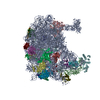 9h3vC  9h3wC  9h3xC  9h3yC  9h3zC  9ha1C  9ha2C  9ha3C  9ha4C  9ha5C  9ha6C  9haiC 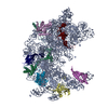 9halC  9hamC M: map data used to model this data C: citing same article ( |
|---|---|
| Similar structure data |


-Links
 PDBj
PDBj


- Assembly
| Deposited unit | 
|
|---|---|
| 1 |
|
-Components
-Large ribosomal subunit protein ... , 15 types, 15 molecules 2FJLNOQRTUVWYZE
| #1: Protein/peptide | Mass: 5397.463 Da / Num. of mol.: 1 / Source method: isolated from a natural source / Source: (natural) |
|---|---|
| #3: Protein | Mass: 20073.234 Da / Num. of mol.: 1 / Source method: isolated from a natural source / Source: (natural) |
| #4: Protein | Mass: 16050.606 Da / Num. of mol.: 1 / Source method: isolated from a natural source / Source: (natural) |
| #5: Protein | Mass: 14877.273 Da / Num. of mol.: 1 / Source method: isolated from a natural source / Source: (natural) |
| #6: Protein | Mass: 13721.938 Da / Num. of mol.: 1 / Source method: isolated from a natural source / Source: (natural) |
| #7: Protein | Mass: 12663.471 Da / Num. of mol.: 1 / Source method: isolated from a natural source / Source: (natural) |
| #8: Protein | Mass: 13396.828 Da / Num. of mol.: 1 / Source method: isolated from a natural source / Source: (natural) |
| #9: Protein | Mass: 11586.374 Da / Num. of mol.: 1 / Source method: isolated from a natural source / Source: (natural) |
| #10: Protein | Mass: 10546.472 Da / Num. of mol.: 1 / Source method: isolated from a natural source / Source: (natural) |
| #11: Protein | Mass: 11078.874 Da / Num. of mol.: 1 / Source method: isolated from a natural source / Source: (natural) |
| #12: Protein | Mass: 10713.465 Da / Num. of mol.: 1 / Source method: isolated from a natural source / Source: (natural) |
| #13: Protein | Mass: 8174.394 Da / Num. of mol.: 1 / Source method: isolated from a natural source / Source: (natural) |
| #14: Protein | Mass: 7286.464 Da / Num. of mol.: 1 / Source method: isolated from a natural source / Source: (natural) |
| #15: Protein | Mass: 6423.625 Da / Num. of mol.: 1 / Source method: isolated from a natural source / Source: (natural) |
| #18: Protein | Mass: 22121.566 Da / Num. of mol.: 1 / Source method: isolated from a natural source / Source: (natural) |
-RNA chain , 2 types, 2 molecules BA
| #2: RNA chain | Mass: 38813.133 Da / Num. of mol.: 1 / Source method: isolated from a natural source / Source: (natural) |
|---|---|
| #16: RNA chain | Mass: 941322.188 Da / Num. of mol.: 1 / Source method: isolated from a natural source / Source: (natural) |
-Protein / Protein/peptide , 2 types, 2 molecules Dy
| #17: Protein | Mass: 22277.535 Da / Num. of mol.: 1 / Source method: isolated from a natural source / Source: (natural) |
|---|---|
| #19: Protein/peptide | Mass: 2084.452 Da / Num. of mol.: 1 / Source method: obtained synthetically / Source: (synth.) |
-Details
| Has protein modification | N |
|---|
-Experimental details
-Experiment
| Experiment | Method: ELECTRON MICROSCOPY |
|---|---|
| EM experiment | Aggregation state: PARTICLE / 3D reconstruction method: single particle reconstruction |
- Sample preparation
| Component | Name: pre50S precursor derived from Api137-treated cells supplemented with Api137 Type: RIBOSOME / Entity ID: all / Source: NATURAL |
|---|---|
| Source (natural) | Organism: |
| Buffer solution | pH: 7.6 |
| Specimen | Embedding applied: NO / Shadowing applied: NO / Staining applied: NO / Vitrification applied: YES |
| Specimen support | Grid material: COPPER / Grid mesh size: 400 divisions/in. / Grid type: Quantifoil |
| Vitrification | Instrument: FEI VITROBOT MARK IV / Cryogen name: ETHANE / Humidity: 95 % / Chamber temperature: 277 K |
- Electron microscopy imaging
| Experimental equipment |  Model: Titan Krios / Image courtesy: FEI Company |
|---|---|
| Microscopy | Model: TFS KRIOS |
| Electron gun | Electron source:  FIELD EMISSION GUN / Accelerating voltage: 300 kV / Illumination mode: OTHER FIELD EMISSION GUN / Accelerating voltage: 300 kV / Illumination mode: OTHER |
| Electron lens | Mode: BRIGHT FIELD / Nominal defocus max: 2000 nm / Nominal defocus min: 800 nm / Cs: 2.7 mm / C2 aperture diameter: 100 µm / Alignment procedure: COMA FREE |
| Image recording | Average exposure time: 1.2 sec. / Electron dose: 46.2 e/Å2 / Film or detector model: GATAN K3 BIOQUANTUM (6k x 4k) / Num. of real images: 2794 |
| EM imaging optics | Energyfilter name: GIF Bioquantum / Energyfilter slit width: 20 eV |
- Processing
| EM software | Name: cryoSPARC / Version: 4.21 / Category: final Euler assignment |
|---|---|
| CTF correction | Type: PHASE FLIPPING AND AMPLITUDE CORRECTION |
| 3D reconstruction | Resolution: 4.37 Å / Resolution method: FSC 0.143 CUT-OFF / Num. of particles: 28649 / Symmetry type: POINT |
