 Movie
Movie Controller

[English] 日本語
 Yorodumi
- PDB-9h48: Mouse Iodothyronine deiodinase 2 catalytic core, mutant - LysLys1... -
+ Open data

- Basic information
| Entry | Database: PDB / ID: 9h48 | ||||||
|---|---|---|---|---|---|---|---|
| Title | Mouse Iodothyronine deiodinase 2 catalytic core, mutant - LysLys180AlaAla, Secys-> Cys | ||||||
 Components Components | Type II iodothyronine deiodinase | ||||||
 Keywords Keywords | OXIDOREDUCTASE / Thioredoxin-fold / iodothyronine deiodinase | ||||||
| Function / homology |  Function and homology information Function and homology informationthyroxine 5'-deiodinase / : / thyroxine 5'-deiodinase activity / thyroid hormone catabolic process / Regulation of thyroid hormone activity / thyroid-stimulating hormone secretion / hormone biosynthetic process / brown fat cell differentiation / positive regulation of cold-induced thermogenesis / plasma membrane Similarity search - Function | ||||||
| Biological species |  | ||||||
| Method |  X-RAY DIFFRACTION / SYNCHROTRON / MOLECULAR REPLACEMENT / Resolution: 1.09 Å X-RAY DIFFRACTION / SYNCHROTRON / MOLECULAR REPLACEMENT / Resolution: 1.09 Å | ||||||
 Authors Authors | Towell, H. / Steegborn, C. | ||||||
| Funding support |  Germany, 1items Germany, 1items
| ||||||
 Citation Citation | Journal: Biomolecules / Year: 2024 Title: Structural Insights into the Iodothyronine Deiodinase 2 Catalytic Core and Deiodinase Catalysis and Dimerization. Authors: Towell, H. / Braun, D. / Brol, A. / di Fonzo, A. / Rijntjes, E. / Kohrle, J. / Schweizer, U. / Steegborn, C. | ||||||
| History |
|
- Structure visualization
| Structure viewer | Molecule: MolmilJmol/JSmol |
|---|
- Downloads & links
-Download
| PDBx/mmCIF format | 9h48.cif.gz | 148.1 KB | Display | PDBx/mmCIF format |
|---|---|---|---|---|
| PDB format | pdb9h48.ent.gz | 97.2 KB | Display | PDB format |
| PDBx/mmJSON format | 9h48.json.gz | Tree view | PDBx/mmJSON format | |
| Others |  Other downloads Other downloads |
-Validation report
| Summary document | 9h48_validation.pdf.gz | 422.4 KB | Display | wwPDB validaton report |
|---|---|---|---|---|
| Full document | 9h48_full_validation.pdf.gz | 423.9 KB | Display | |
| Data in XML | 9h48_validation.xml.gz | 12.4 KB | Display | |
| Data in CIF | 9h48_validation.cif.gz | 17.4 KB | Display | |
| Arichive directory | https://data.pdbj.org/pub/pdb/validation_reports/h4/9h48ftp://data.pdbj.org/pub/pdb/validation_reports/h4/9h48 | HTTPS FTP |
-Related structure data
| Related structure data | 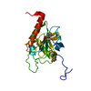 4tr3S S: Starting model for refinement |
|---|---|
| Similar structure data |
-Links
 PDBj
PDBj- Assembly
| Deposited unit | 
| ||||||||||||
|---|---|---|---|---|---|---|---|---|---|---|---|---|---|
| 1 |
| ||||||||||||
| Unit cell |
|
-Components
| #1: Protein | Mass: 21347.764 Da / Num. of mol.: 1 / Mutation: K180A, K181A, U126C Source method: isolated from a genetically manipulated source Source: (gene. exp.)  |
|---|---|
| #2: Water | ChemComp-HOH /  Mass: 18.015 Da / Num. of mol.: 192 / Source method: isolated from a natural source / Formula: H2O Mass: 18.015 Da / Num. of mol.: 192 / Source method: isolated from a natural source / Formula: H2O |
| Has protein modification | N |
-Experimental details
-Experiment
| Experiment | Method: X-RAY DIFFRACTION / Number of used crystals: 1 |
|---|
- Sample preparation
| Crystal | Density Matthews: 2 Å3/Da / Density % sol: 38.38 % |
|---|---|
| Crystal grow | Temperature: 277 K / Method: vapor diffusion, sitting drop / Details: 0.1M SPG buffer pH 7.0, 25% (w/v) PEG 1500 |
-Data collection
| Diffraction | Mean temperature: 100 K / Serial crystal experiment: N |
|---|---|
| Diffraction source | Source: SYNCHROTRON / Site: BESSY / Beamline: 14.1 / Wavelength: 0.9184 Å |
| Detector | Type: DECTRIS PILATUS3 6M / Detector: PIXEL / Date: Feb 27, 2020 |
| Radiation | Protocol: SINGLE WAVELENGTH / Monochromatic (M) / Laue (L): M / Scattering type: x-ray |
| Radiation wavelength | Wavelength: 0.9184 Å / Relative weight: 1 |
| Reflection | Resolution: 1.089→34.85 Å / Num. obs: 65565 / % possible obs: 94.85 % / Redundancy: 5.4 % / Biso Wilson estimate: 14.23 Å2 / CC1/2: 0.999 / Rmerge(I) obs: 0.0694 / Rrim(I) all: 0.07659 / Net I/σ(I): 9.45 |
| Reflection shell | Resolution: 1.089→1.128 Å / Redundancy: 3.6 % / Mean I/σ(I) obs: 0.39 / Num. unique obs: 4435 / CC1/2: 0.117 / % possible all: 64.62 |
- Processing
| Software |
| ||||||||||||||||||||||||
|---|---|---|---|---|---|---|---|---|---|---|---|---|---|---|---|---|---|---|---|---|---|---|---|---|---|
| Refinement | Method to determine structure: MOLECULAR REPLACEMENT Starting model: 4TR3 Resolution: 1.09→34.85 Å / Cross valid method: FREE R-VALUE Stereochemistry target values: GeoStd + Monomer Library + CDL v1.2
| ||||||||||||||||||||||||
| Solvent computation | Solvent model: FLAT BULK SOLVENT MODEL | ||||||||||||||||||||||||
| Displacement parameters | Biso mean: 21.19 Å2 | ||||||||||||||||||||||||
| Refinement step | Cycle: LAST / Resolution: 1.09→34.85 Å
| ||||||||||||||||||||||||
| Refine LS restraints |
| ||||||||||||||||||||||||
| LS refinement shell | Resolution: 1.09→1.11 Å
|
