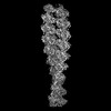
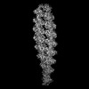
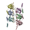
Journal: Nucleic Acids Res / Year: 2025 Title: Structural basis for cooperative ssDNA binding by bacteriophage protein filament P12. Authors: Lena K Träger / Morris Degen / Joana Pereira / Janani Durairaj / Raphael Dias Teixeira / Sebastian Hiller / Nicolas Huguenin-Dezot / Abstract: Protein-primed DNA replication is a unique mechanism, bioorthogonal to other known DNA replication modes. It relies on specialised single-stranded DNA (ssDNA)-binding proteins (SSBs) to stabilise ...Protein-primed DNA replication is a unique mechanism, bioorthogonal to other known DNA replication modes. It relies on specialised single-stranded DNA (ssDNA)-binding proteins (SSBs) to stabilise ssDNA intermediates by unknown mechanisms. Here, we present the structural and biochemical characterisation of P12, an SSB from bacteriophage PRD1. High-resolution cryo-electron microscopy reveals that P12 forms a unique, cooperative filament along ssDNA. Each protomer binds the phosphate backbone of 6 nucleotides in a sequence-independent manner, protecting ssDNA from nuclease degradation. Filament formation is driven by an intrinsically disordered C-terminal tail, facilitating cooperative binding. We identify residues essential for ssDNA interaction and link the ssDNA-binding ability of P12 to toxicity in host cells. Bioinformatic analyses place the P12 fold as a distinct branch within the OB-like fold family. This work offers new insights into protein-primed DNA replication and lays a foundation for biotechnological applications.
In the structure databanks used in Yorodumi, some data are registered as the other names, "COVID-19 virus" and "2019-nCoV". Here are the details of the virus and the list of structure data.
Jan 31, 2019. EMDB accession codes are about to change! (news from PDBe EMDB page)
EMDB accession codes are about to change! (news from PDBe EMDB page)
The allocation of 4 digits for EMDB accession codes will soon come to an end. Whilst these codes will remain in use, new EMDB accession codes will include an additional digit and will expand incrementally as the available range of codes is exhausted. The current 4-digit format prefixed with “EMD-” (i.e. EMD-XXXX) will advance to a 5-digit format (i.e. EMD-XXXXX), and so on. It is currently estimated that the 4-digit codes will be depleted around Spring 2019, at which point the 5-digit format will come into force.
The EM Navigator/Yorodumi systems omit the EMD- prefix.
Related info.:Q: What is EMD? / ID/Accession-code notation in Yorodumi/EM Navigator
Yorodumi is a browser for structure data from EMDB, PDB, SASBDB, etc.
This page is also the successor to EM Navigator detail page, and also detail information page/front-end page for Omokage search.
The word "yorodu" (or yorozu) is an old Japanese word meaning "ten thousand". "mi" (miru) is to see.
Related info.:EMDB / PDB / SASBDB / Comparison of 3 databanks / Yorodumi Search / Aug 31, 2016. New EM Navigator & Yorodumi / Yorodumi Papers / Jmol/JSmol / Function and homology information / Changes in new EM Navigator and Yorodumi
 Movie
Movie Controller


 Open data
 Basic information
 Components
 Keywords
 Function and homology information
 Enterobacteria phage PRD1 (virus)
 X-RAY DIFFRACTION /
  Authors
 Switzerland, 1items
  Citation
 Structure visualization
 Downloads & links
 Other downloads
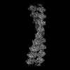
 PDBj
PDBj Assembly

 Mass: 18.015 Da / Num. of mol.: 58 / Source method: isolated from a natural source / Formula: H2O
 Sample preparation
 Processing