 Movie
Movie Controller

[English] 日本語
 Yorodumi
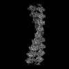
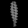
- EMDB-52709: Enterobacteriaphage PRD1 - P12 protein filament in complex with r... -
+ Open data

- Basic information
| Entry | 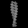 | |||||||||
|---|---|---|---|---|---|---|---|---|---|---|
| Title | Enterobacteriaphage PRD1 - P12 protein filament in complex with repetitive (ATGCT) ssDNA | |||||||||
 Map data Map data | Primary Map | |||||||||
 Sample Sample |
| |||||||||
 Keywords Keywords | SSB / Protein primed replication / PRD1 / Protein filament / Homooligomer / ssDNA-binding / REPLICATION | |||||||||
| Function / homology | nucleotide binding / DNA binding / Single-stranded DNA-binding protein Function and homology information Function and homology information | |||||||||
| Biological species |   Enterobacteria phage PRD1 (virus) / synthetic construct (others) Enterobacteria phage PRD1 (virus) / synthetic construct (others) | |||||||||
| Method | helical reconstruction / cryo EM / Resolution: 3.11 Å | |||||||||
 Authors Authors | Degen M / Traeger KL / Hiller S | |||||||||
| Funding support |  Switzerland, 1 items Switzerland, 1 items
| |||||||||
 Citation Citation | Journal: Nucleic Acids Res / Year: 2025 Title: Structural basis for cooperative ssDNA binding by bacteriophage protein filament P12. Authors: Lena K Träger / Morris Degen / Joana Pereira / Janani Durairaj / Raphael Dias Teixeira / Sebastian Hiller / Nicolas Huguenin-Dezot / Abstract: Protein-primed DNA replication is a unique mechanism, bioorthogonal to other known DNA replication modes. It relies on specialised single-stranded DNA (ssDNA)-binding proteins (SSBs) to stabilise ...Protein-primed DNA replication is a unique mechanism, bioorthogonal to other known DNA replication modes. It relies on specialised single-stranded DNA (ssDNA)-binding proteins (SSBs) to stabilise ssDNA intermediates by unknown mechanisms. Here, we present the structural and biochemical characterisation of P12, an SSB from bacteriophage PRD1. High-resolution cryo-electron microscopy reveals that P12 forms a unique, cooperative filament along ssDNA. Each protomer binds the phosphate backbone of 6 nucleotides in a sequence-independent manner, protecting ssDNA from nuclease degradation. Filament formation is driven by an intrinsically disordered C-terminal tail, facilitating cooperative binding. We identify residues essential for ssDNA interaction and link the ssDNA-binding ability of P12 to toxicity in host cells. Bioinformatic analyses place the P12 fold as a distinct branch within the OB-like fold family. This work offers new insights into protein-primed DNA replication and lays a foundation for biotechnological applications. | |||||||||
| History |
|
- Structure visualization
| Supplemental images |
|---|

- Downloads & links
-EMDB archive
| Map data | emd_52709.map.gz | 168 MB | EMDB map data format | |
|---|---|---|---|---|
| Header (meta data) | emd-52709-v30.xmlemd-52709.xml | 17.2 KB 17.2 KB | Display Display | EMDB header |
| FSC (resolution estimation) | emd_52709_fsc.xml | 11.9 KB | Display | FSC data file |
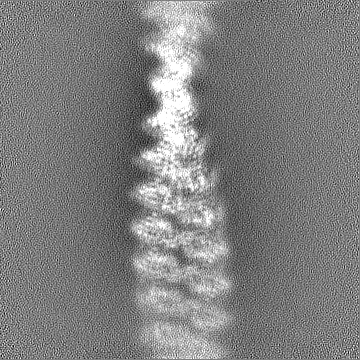
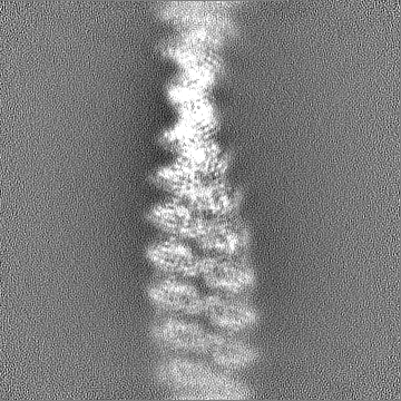
| Images |  emd_52709.png emd_52709.png | 50.8 KB | ||
| Filedesc metadata | emd-52709.cif.gz | 4.9 KB | ||
| Others | emd_52709_additional_1.map.gzemd_52709_half_map_1.map.gzemd_52709_half_map_2.map.gz | 148.4 MB 165 MB 165 MB | ||
| Archive directory |  http://ftp.pdbj.org/pub/emdb/structures/EMD-52709ftp://ftp.pdbj.org/pub/emdb/structures/EMD-52709 http://ftp.pdbj.org/pub/emdb/structures/EMD-52709ftp://ftp.pdbj.org/pub/emdb/structures/EMD-52709 | HTTPS FTP |
-Related structure data
| Related structure data | 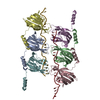 9i86MC  9gfqC C: citing same article ( M: atomic model generated by this map |
|---|---|
| Similar structure data |
-Links
| EMDB pages | EMDB (EBI/PDBe) / EMDataResource |
|---|
-Map
| File | Download / File: emd_52709.map.gz / Format: CCP4 / Size: 178 MB / Type: IMAGE STORED AS FLOATING POINT NUMBER (4 BYTES) | ||||||||||||||||||||||||||||||||||||
|---|---|---|---|---|---|---|---|---|---|---|---|---|---|---|---|---|---|---|---|---|---|---|---|---|---|---|---|---|---|---|---|---|---|---|---|---|---|
| Annotation | Primary Map | ||||||||||||||||||||||||||||||||||||
| Projections & slices | Image control
Images are generated by Spider. | ||||||||||||||||||||||||||||||||||||
| Voxel size | X=Y=Z: 0.82 Å | ||||||||||||||||||||||||||||||||||||
| Density |
| ||||||||||||||||||||||||||||||||||||
| Symmetry | Space group: 1 | ||||||||||||||||||||||||||||||||||||
| Details | EMDB XML:
|
 Z (Sec.)
 Y (Row.)
 X (Col.)


-Supplemental data
-Additional map: DeepEMhancer Map
| File | emd_52709_additional_1.map | ||||||||||||
|---|---|---|---|---|---|---|---|---|---|---|---|---|---|
| Annotation | DeepEMhancer Map | ||||||||||||
| Projections & Slices |
| ||||||||||||
| Density Histograms |


-Half map: Half map A
| File | emd_52709_half_map_1.map | ||||||||||||
|---|---|---|---|---|---|---|---|---|---|---|---|---|---|
| Annotation | Half map A | ||||||||||||
| Projections & Slices |
| ||||||||||||
| Density Histograms |


-Half map: Half Map B
| File | emd_52709_half_map_2.map | ||||||||||||
|---|---|---|---|---|---|---|---|---|---|---|---|---|---|
| Annotation | Half Map B | ||||||||||||
| Projections & Slices |
| ||||||||||||
| Density Histograms |


- Sample components
-Entire : P12 filament bound to ssDNA
| Entire | Name: P12 filament bound to ssDNA |
|---|---|
| Components |
|
-Supramolecule #1: P12 filament bound to ssDNA
| Supramolecule | Name: P12 filament bound to ssDNA / type: complex / ID: 1 / Parent: 0 / Macromolecule list: all |
|---|---|
| Source (natural) | Organism: Enterobacteria phage PRD1 (virus) |
-Macromolecule #1: Enterobacteriaphage PRD1 - P12 protein filament
| Macromolecule | Name: Enterobacteriaphage PRD1 - P12 protein filament / type: protein_or_peptide / ID: 1 / Enantiomer: LEVO |
|---|---|
| Source (natural) | Organism: Enterobacteria phage PRD1 (virus) |
| Recombinant expression | Organism:  |
| Sequence | String: MEIVSKLTLK TIGAQPKPHS VKENTALASI YGRVRGKKVG QSTFGDFIKF EGEFEGVNIA TGEVFRSGAL ILPKVLESLL AGAVDGENTV DFAVEIWAKP SEKGNTGYEY GVKPLIEPAA SDELAALRNQ VKAALPAPAA AGEAAAEAKP AAKAKAKAEA UniProtKB: Single-stranded DNA-binding protein |
-Macromolecule #2: ssDNA repetitive (ATGCT)x16
| Macromolecule | Name: ssDNA repetitive (ATGCT)x16 / type: dna / ID: 2 / Classification: DNA |
|---|---|
| Source (natural) | Organism: synthetic construct (others) |
| Sequence | String: ATGCTATGCT ATGCTATGCT ATGCTATGCT ATGCTATGCT ATGCTATGCT ATGCTATGCT ATGCTATGCT ATGCTATGCT |
-Experimental details
-Structure determination
| Method | cryo EM |
|---|---|
 Processing Processing | helical reconstruction |
| Aggregation state | filament |
-Sample preparation
| Buffer | pH: 8.5 |
|---|---|
| Vitrification | Cryogen name: ETHANE |
- Electron microscopy
| Microscope | TFS KRIOS |
|---|---|
| Image recording | Film or detector model: GATAN K2 SUMMIT (4k x 4k) / Average electron dose: 49.41 e/Å2 |
| Electron beam | Acceleration voltage: 300 kV / Electron source:  FIELD EMISSION GUN FIELD EMISSION GUN |
| Electron optics | Illumination mode: FLOOD BEAM / Imaging mode: BRIGHT FIELD / Cs: 2.7 mm / Nominal defocus max: 1.8 µm / Nominal defocus min: 0.11 µm |
| Experimental equipment |  Model: Titan Krios / Image courtesy: FEI Company |
-Image processing
| Final reconstruction | Applied symmetry - Helical parameters - Δz: 28.281 Å Applied symmetry - Helical parameters - Δ&Phi: 17.969 ° Applied symmetry - Helical parameters - Axial symmetry: C1 (asymmetric) Resolution.type: BY AUTHOR / Resolution: 3.11 Å / Resolution method: FSC 0.143 CUT-OFF / Software - Name: cryoSPARC (ver. 4.4.1) / Number images used: 183109 |
|---|---|
| Startup model | Type of model: PDB ENTRY PDB model - PDB ID: |
| Final angle assignment | Type: NOT APPLICABLE / Software - Name: cryoSPARC (ver. 4.4.1) |
| FSC plot (resolution estimation) |  |
