 Movie
Movie Controller

[English] 日本語
 Yorodumi
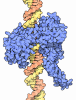
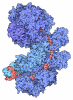
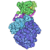
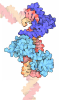
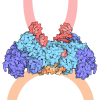
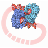
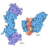
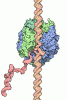
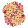
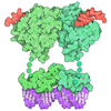
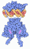
- PDB-9gbv: E.coli gyrase holocomplex with chirally wrapped 217 bp DNA fragment -
+ Open data

- Basic information
| Entry | Database: PDB / ID: 9gbv | |||||||||||||||
|---|---|---|---|---|---|---|---|---|---|---|---|---|---|---|---|---|
| Title | E.coli gyrase holocomplex with chirally wrapped 217 bp DNA fragment | |||||||||||||||
 Components Components |
| |||||||||||||||
 Keywords Keywords | ISOMERASE / DNA gyrase / type II topoisomerase / supercoiling / DNA crossover | |||||||||||||||
| Function / homology |  Function and homology information Function and homology informationAction of antimicrobials / DNA topoisomerase type II (double strand cut, ATP-hydrolyzing) complex / DNA negative supercoiling activity / DNA topoisomerase type II (double strand cut, ATP-hydrolyzing) activity / DNA topoisomerase (ATP-hydrolysing) / Antimicrobial resistance / negative regulation of DNA-templated DNA replication / DNA topological change / ATP-dependent activity, acting on DNA / DNA-templated DNA replication ...Action of antimicrobials / DNA topoisomerase type II (double strand cut, ATP-hydrolyzing) complex / DNA negative supercoiling activity / DNA topoisomerase type II (double strand cut, ATP-hydrolyzing) activity / DNA topoisomerase (ATP-hydrolysing) / Antimicrobial resistance / negative regulation of DNA-templated DNA replication / DNA topological change / ATP-dependent activity, acting on DNA / DNA-templated DNA replication / response to xenobiotic stimulus / chromosome / DNA-templated transcription / DNA binding / ATP binding / membrane / identical protein binding / cytosol / cytoplasm Similarity search - Function | |||||||||||||||
| Biological species |   Escherichia coli (E. coli) Escherichia coli (E. coli) Escherichia phage Mu (virus) Escherichia phage Mu (virus) | |||||||||||||||
| Method | ELECTRON MICROSCOPY / single particle reconstruction / cryo EM / Resolution: 2.32 Å | |||||||||||||||
 Authors Authors | Michalczyk, E. / Ghilarov, D. | |||||||||||||||
| Funding support |  United Kingdom, United Kingdom,  Poland, 3items Poland, 3items
| |||||||||||||||
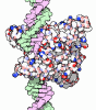
 Citation Citation | Journal: Proc Natl Acad Sci U S A / Year: 2024 Title: Structural basis of chiral wrap and T-segment capture by DNA gyrase. Authors: Elizabeth Michalczyk / Zuzanna Pakosz-Stępień / Jonathon D Liston / Olivia Gittins / Marta Pabis / Jonathan G Heddle / Dmitry Ghilarov / Abstract: Type II topoisomerase DNA gyrase transduces the energy of ATP hydrolysis into the negative supercoiling of DNA. The postulated catalytic mechanism involves stabilization of a chiral DNA loop followed ...Type II topoisomerase DNA gyrase transduces the energy of ATP hydrolysis into the negative supercoiling of DNA. The postulated catalytic mechanism involves stabilization of a chiral DNA loop followed by the passage of the T-segment through the temporarily cleaved G-segment resulting in sign inversion. The molecular basis for this is poorly understood as the chiral loop has never been directly observed. We have obtained high-resolution cryoEM structures of gyrase with chirally wrapped 217 bp DNA with and without the fluoroquinolone moxifloxacin (MFX). Each structure constrains a positively supercoiled figure-of-eight DNA loop stabilized by a GyrA β-pinwheel domain which has the structure of a flat disc. By comparing the catalytic site of the native drug-free and MFX-bound gyrase structures both of which contain a single metal ion, we demonstrate that the enzyme is observed in a native precatalytic state. Our data imply that T-segment trapping is not dependent on the dimerization of the ATPase domains which appears to only be possible after strand passage has taken place. | |||||||||||||||
| History |
|
- Structure visualization
| Structure viewer | Molecule: MolmilJmol/JSmol |
|---|
- Downloads & links
-Download
| PDBx/mmCIF format | 9gbv.cif.gz | 786.2 KB | Display | PDBx/mmCIF format |
|---|---|---|---|---|
| PDB format | pdb9gbv.ent.gz | 624.5 KB | Display | PDB format |
| PDBx/mmJSON format | 9gbv.json.gz | Tree view | PDBx/mmJSON format | |
| Others |  Other downloads Other downloads |
-Validation report
| Arichive directory | https://data.pdbj.org/pub/pdb/validation_reports/gb/9gbvftp://data.pdbj.org/pub/pdb/validation_reports/gb/9gbv | HTTPS FTP |
|---|
-Related structure data
| Related structure data |  51222MC 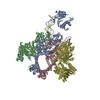 9ggqC M: map data used to model this data C: citing same article ( |
|---|---|
| Similar structure data |
-Links
 PDBj
PDBj










- Assembly
| Deposited unit | 
|
|---|---|
| 1 |
|
-Components
| #1: Protein | Mass: 97646.195 Da / Num. of mol.: 2 Source method: isolated from a genetically manipulated source Source: (gene. exp.) Escherichia coli (E. coli) / Gene: gyrA, hisW, nalA, parD, b2231, JW2225 / Production host: Escherichia coli BL21(DE3) (bacteria)References: UniProt: P0AES4, DNA topoisomerase (ATP-hydrolysing) #2: Protein | Mass: 90891.734 Da / Num. of mol.: 2 Source method: isolated from a genetically manipulated source Source: (gene. exp.) Escherichia coli (E. coli)Gene: gyrB, acrB, cou, himB, hisU, nalC, parA, pcbA, b3699, JW5625 Production host: Escherichia coli (E. coli)References: UniProt: P0AES6, DNA topoisomerase (ATP-hydrolysing) #3: DNA chain | | Mass: 65926.109 Da / Num. of mol.: 1 Source method: isolated from a genetically manipulated source Source: (gene. exp.) Escherichia phage Mu (virus) / Production host: Escherichia coli (E. coli) / References: GenBank: 215533#4: DNA chain | | Mass: 66202.258 Da / Num. of mol.: 1 Source method: isolated from a genetically manipulated source Source: (gene. exp.) Escherichia phage Mu (virus) / Production host: Escherichia coli (E. coli) / References: GenBank: 215533#5: Chemical |   Mass: 24.305 Da / Num. of mol.: 2 / Source method: obtained synthetically / Formula: Mg Mass: 24.305 Da / Num. of mol.: 2 / Source method: obtained synthetically / Formula: MgHas ligand of interest | N | Has protein modification | N | |
|---|
-Experimental details
-Experiment
| Experiment | Method: ELECTRON MICROSCOPY |
|---|---|
| EM experiment | Aggregation state: PARTICLE / 3D reconstruction method: single particle reconstruction |
- Sample preparation
| Component |
| ||||||||||||||||||||||||
|---|---|---|---|---|---|---|---|---|---|---|---|---|---|---|---|---|---|---|---|---|---|---|---|---|---|
| Molecular weight | Experimental value: NO | ||||||||||||||||||||||||
| Source (natural) |
| ||||||||||||||||||||||||
| Source (recombinant) |
| ||||||||||||||||||||||||
| Buffer solution | pH: 8 | ||||||||||||||||||||||||
| Specimen | Embedding applied: NO / Shadowing applied: NO / Staining applied: NO / Vitrification applied: YES | ||||||||||||||||||||||||
| Vitrification | Instrument: FEI VITROBOT MARK IV / Cryogen name: ETHANE / Humidity: 95 % / Chamber temperature: 283 K |
- Electron microscopy imaging
| Experimental equipment |  Model: Titan Krios / Image courtesy: FEI Company |
|---|---|
| Microscopy | Model: FEI TITAN KRIOS |
| Electron gun | Electron source:  FIELD EMISSION GUN / Accelerating voltage: 300 kV / Illumination mode: FLOOD BEAM FIELD EMISSION GUN / Accelerating voltage: 300 kV / Illumination mode: FLOOD BEAM |
| Electron lens | Mode: BRIGHT FIELD / Nominal defocus max: 2100 nm / Nominal defocus min: 900 nm |
| Image recording | Electron dose: 41.84 e/Å2 / Film or detector model: GATAN K3 BIOQUANTUM (6k x 4k) |
- Processing
| EM software |
| ||||||||||||||||||||||||||||
|---|---|---|---|---|---|---|---|---|---|---|---|---|---|---|---|---|---|---|---|---|---|---|---|---|---|---|---|---|---|
| CTF correction | Type: PHASE FLIPPING AND AMPLITUDE CORRECTION | ||||||||||||||||||||||||||||
| Symmetry | Point symmetry: C1 (asymmetric) | ||||||||||||||||||||||||||||
| 3D reconstruction | Resolution: 2.32 Å / Resolution method: FSC 0.143 CUT-OFF / Num. of particles: 170369 / Symmetry type: POINT | ||||||||||||||||||||||||||||
| Atomic model building | Protocol: AB INITIO MODEL |
