 Movie
Movie Controller

[English] 日本語
 Yorodumi
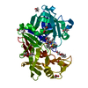
- PDB-9g35: The HIV protease inhibitor lopinavir binding to the active site o... -
+ Open data

- Basic information
| Entry | Database: PDB / ID: 9g35 | |||||||||
|---|---|---|---|---|---|---|---|---|---|---|
| Title | The HIV protease inhibitor lopinavir binding to the active site of Cryphonectria parasitica endothiapepsin | |||||||||
 Components Components | Endothiapepsin | |||||||||
 Keywords Keywords | HYDROLASE / aspartic protease / drug development / inhibitor / X-ray crystallographic screening / chestnut blight fungus / protease model | |||||||||
| Function / homology |  Function and homology information Function and homology information | |||||||||
| Biological species |  Cryphonectria parasitica (chestnut blight fungus) Cryphonectria parasitica (chestnut blight fungus) | |||||||||
| Method |  X-RAY DIFFRACTION / SYNCHROTRON / MOLECULAR REPLACEMENT / Resolution: 1.5 Å X-RAY DIFFRACTION / SYNCHROTRON / MOLECULAR REPLACEMENT / Resolution: 1.5 Å | |||||||||
 Authors Authors | Falke, S. / Senst, J.M. / Guenther, S. / Meents, A. | |||||||||
| Funding support |  Germany, 2items Germany, 2items
| |||||||||
 Citation Citation | Journal: To Be Published Title: The HIV protease inhibitor lopinavir binding to the active site of Cryphonectria parasitica endothiapepsin Authors: Falke, S. / Senst, J.M. / Guenther, S. / Meents, A. | |||||||||
| History |
|
- Structure visualization
| Structure viewer | Molecule: MolmilJmol/JSmol |
|---|
- Downloads & links
-Download
| PDBx/mmCIF format | 9g35.cif.gz | 105.5 KB | Display | PDBx/mmCIF format |
|---|---|---|---|---|
| PDB format | pdb9g35.ent.gz | 62.6 KB | Display | PDB format |
| PDBx/mmJSON format | 9g35.json.gz | Tree view | PDBx/mmJSON format | |
| Others |  Other downloads Other downloads |
-Validation report
| Arichive directory | https://data.pdbj.org/pub/pdb/validation_reports/g3/9g35ftp://data.pdbj.org/pub/pdb/validation_reports/g3/9g35 | HTTPS FTP |
|---|
-Related structure data
| Related structure data | |
|---|---|
| Similar structure data |
-Links
 PDBj
PDBj
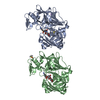
- Assembly
| Deposited unit | 
| ||||||||||||
|---|---|---|---|---|---|---|---|---|---|---|---|---|---|
| 1 |
| ||||||||||||
| Unit cell |
|
-Components
-Protein , 1 types, 1 molecules A
| #1: Protein | Mass: 33813.855 Da / Num. of mol.: 1 / Source method: isolated from a natural source Source: (natural) Cryphonectria parasitica (chestnut blight fungus)References: UniProt: P11838, endothiapepsin |
|---|
-Non-polymers , 5 types, 393 molecules 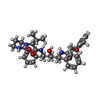


| #2: Chemical | ChemComp-AB1 /  Mass: 628.801 Da / Num. of mol.: 1 / Source method: obtained synthetically / Formula: C37H48N4O5 / Feature type: SUBJECT OF INVESTIGATION Mass: 628.801 Da / Num. of mol.: 1 / Source method: obtained synthetically / Formula: C37H48N4O5 / Feature type: SUBJECT OF INVESTIGATION | ||||
|---|---|---|---|---|---|
| #3: Chemical | ChemComp-PGE /  Mass: 150.173 Da / Num. of mol.: 1 / Source method: obtained synthetically / Formula: C6H14O4 Mass: 150.173 Da / Num. of mol.: 1 / Source method: obtained synthetically / Formula: C6H14O4 | ||||
| #4: Chemical |  Mass: 62.068 Da / Num. of mol.: 2 / Source method: obtained synthetically / Formula: C2H6O2 Mass: 62.068 Da / Num. of mol.: 2 / Source method: obtained synthetically / Formula: C2H6O2#5: Chemical |  Mass: 194.226 Da / Num. of mol.: 2 / Source method: obtained synthetically / Formula: C8H18O5 / Comment: precipitant*YM Mass: 194.226 Da / Num. of mol.: 2 / Source method: obtained synthetically / Formula: C8H18O5 / Comment: precipitant*YM#6: Water | ChemComp-HOH / | Mass: 18.015 Da / Num. of mol.: 387 / Source method: isolated from a natural source / Formula: H2O |
-Details
| Has ligand of interest | Y |
|---|---|
| Has protein modification | Y |
-Experimental details
-Experiment
| Experiment | Method: X-RAY DIFFRACTION / Number of used crystals: 1 |
|---|
- Sample preparation
| Crystal | Density Matthews: 2.48 Å3/Da / Density % sol: 50.45 % |
|---|---|
| Crystal grow | Temperature: 293 K / Method: vapor diffusion, hanging drop Details: 24% (w/v) PEG 4000, 0.1 M ammonium acetate and 0.1 M sodium acetate pH 4.6 were mixed with an equal volume of 6 mg/ml endothiapepsin solution. Microseeding was applied. After 2 days crystals ...Details: 24% (w/v) PEG 4000, 0.1 M ammonium acetate and 0.1 M sodium acetate pH 4.6 were mixed with an equal volume of 6 mg/ml endothiapepsin solution. Microseeding was applied. After 2 days crystals were soaked with the ligand in reservoir solution in the presence of 5% (v/v) DMSO. |
-Data collection
| Diffraction | Mean temperature: 100 K / Serial crystal experiment: N |
|---|---|
| Diffraction source | Source: SYNCHROTRON / Site: PETRA III, DESY / Beamline: P11 / Wavelength: 1.03319 Å |
| Detector | Type: DECTRIS EIGER2 X 16M / Detector: PIXEL / Date: May 3, 2024 |
| Radiation | Protocol: SINGLE WAVELENGTH / Monochromatic (M) / Laue (L): M / Scattering type: x-ray |
| Radiation wavelength | Wavelength: 1.03319 Å / Relative weight: 1 |
| Reflection | Resolution: 1.5→42.75 Å / Num. obs: 101583 / % possible obs: 97.4 % / Redundancy: 3.4 % / Biso Wilson estimate: 11.07 Å2 / CC1/2: 0.998 / Rrim(I) all: 0.047 / Net I/σ(I): 20.77 |
| Reflection shell | Resolution: 1.5→1.54 Å / Num. unique obs: 7716 / CC1/2: 0.987 / Rrim(I) all: 0.113 |
- Processing
| Software |
| |||||||||||||||||||||||||||||||||||||||||||||||||||||||||||||||||||||||||||||||||||||||||||||||||||||||||
|---|---|---|---|---|---|---|---|---|---|---|---|---|---|---|---|---|---|---|---|---|---|---|---|---|---|---|---|---|---|---|---|---|---|---|---|---|---|---|---|---|---|---|---|---|---|---|---|---|---|---|---|---|---|---|---|---|---|---|---|---|---|---|---|---|---|---|---|---|---|---|---|---|---|---|---|---|---|---|---|---|---|---|---|---|---|---|---|---|---|---|---|---|---|---|---|---|---|---|---|---|---|---|---|---|---|---|
| Refinement | Method to determine structure: MOLECULAR REPLACEMENT / Resolution: 1.5→42.75 Å / SU ML: 0.1167 / Cross valid method: FREE R-VALUE / σ(F): 1.42 / Phase error: 14.5396 Stereochemistry target values: GeoStd + Monomer Library + CDL v1.2
| |||||||||||||||||||||||||||||||||||||||||||||||||||||||||||||||||||||||||||||||||||||||||||||||||||||||||
| Solvent computation | Shrinkage radii: 0.9 Å / VDW probe radii: 1.11 Å / Solvent model: FLAT BULK SOLVENT MODEL | |||||||||||||||||||||||||||||||||||||||||||||||||||||||||||||||||||||||||||||||||||||||||||||||||||||||||
| Displacement parameters | Biso mean: 13.49 Å2 | |||||||||||||||||||||||||||||||||||||||||||||||||||||||||||||||||||||||||||||||||||||||||||||||||||||||||
| Refinement step | Cycle: LAST / Resolution: 1.5→42.75 Å
| |||||||||||||||||||||||||||||||||||||||||||||||||||||||||||||||||||||||||||||||||||||||||||||||||||||||||
| Refine LS restraints |
| |||||||||||||||||||||||||||||||||||||||||||||||||||||||||||||||||||||||||||||||||||||||||||||||||||||||||
| LS refinement shell |
|
