 Movie
Movie Controller


+ Open data

- Basic information
| Entry | Database: PDB / ID: 9g2r | ||||||
|---|---|---|---|---|---|---|---|
| Title | Endophilin B1 dimers bound to nanodiscs | ||||||
 Components Components | Endophilin-B1 | ||||||
 Keywords Keywords | APOPTOSIS / BAR / N-BAR / endophilin / membrane / curvature / cardiolipin / MSP2N2 / nanodisc / peripheral membrane protein | ||||||
| Function / homology |  Function and homology information Function and homology informationlysophosphatidic acid acyltransferase activity / positive regulation of membrane tubulation / autophagic cell death / protein localization to vacuolar membrane / positive regulation of autophagosome assembly / phosphatidylinositol 3-kinase activator activity / receptor catabolic process / membrane fission / membrane organization / : ...lysophosphatidic acid acyltransferase activity / positive regulation of membrane tubulation / autophagic cell death / protein localization to vacuolar membrane / positive regulation of autophagosome assembly / phosphatidylinositol 3-kinase activator activity / receptor catabolic process / membrane fission / membrane organization / : / autophagosome membrane / cellular response to glucose starvation / regulation of macroautophagy / positive regulation of autophagy / fatty acid binding / cellular response to amino acid starvation / regulation of cytokinesis / regulation of protein stability / positive regulation of protein-containing complex assembly / autophagy / endocytosis / cytoplasmic vesicle / midbody / protein-macromolecule adaptor activity / mitochondrial outer membrane / cadherin binding / Golgi membrane / apoptotic process / protein homodimerization activity / protein-containing complex / membrane / cytosol / cytoplasm Similarity search - Function | ||||||
| Biological species |  Homo sapiens (human) Homo sapiens (human) | ||||||
| Method | ELECTRON MICROSCOPY / single particle reconstruction / cryo EM / Resolution: 3.88 Å | ||||||
 Authors Authors | Thorlacius, A. / Sundborger-Lunna, A. | ||||||
| Funding support |  Sweden, 1items Sweden, 1items
| ||||||
 Citation Citation | Journal: Commun Biol / Year: 2025 Title: Peripheral membrane protein endophilin B1 probes, perturbs and permeabilizes lipid bilayers. Authors: Arni Thorlacius / Maksim Rulev / Oscar Sundberg / Anna Sundborger-Lunna / Abstract: Bin/Amphiphysin/Rvs167 (BAR) domain containing proteins are peripheral membrane proteins that regulate intracellular membrane curvature. BAR protein endophilin B1 plays a key role in multiple ...Bin/Amphiphysin/Rvs167 (BAR) domain containing proteins are peripheral membrane proteins that regulate intracellular membrane curvature. BAR protein endophilin B1 plays a key role in multiple cellular processes critical for oncogenesis, including autophagy and apoptosis. Amphipathic regions in endophilin B1 drive membrane association and tubulation through membrane scaffolding. Our understanding of exactly how BAR proteins like endophilin B1 promote highly diverse intracellular membrane remodeling events in the cell is severely limited due to lack of high-resolution structural information. Here we present the highest resolution cryo-EM structure of a BAR protein to date and the first structures of a BAR protein bound to a lipid bicelle. Using neural networks, we can effectively sort particle species of different stoichiometries, revealing the tremendous flexibility of post-membrane binding, pre-polymer BAR dimer organization and membrane deformation. We also show that endophilin B1 efficiently permeabilizes negatively charged liposomes that contain mitochondria-specific lipid cardiolipin and propose a new model for Bax-mediated cell death. #1: Journal: Biorxiv / Year: 2024Title: Peripheral membrane protein endophilin B1 probes, perturbs and permeabilizes lipid bilayers Authors: Thorlacius, A. / Rulev, M. / Sundberg, O. / Sundborger-Lunna, A. | ||||||
| History |
|
- Structure visualization
| Structure viewer | Molecule: MolmilJmol/JSmol |
|---|
- Downloads & links
-Download
| PDBx/mmCIF format | 9g2r.cif.gz | 496.6 KB | Display | PDBx/mmCIF format |
|---|---|---|---|---|
| PDB format | pdb9g2r.ent.gz | 411.8 KB | Display | PDB format |
| PDBx/mmJSON format | 9g2r.json.gz | Tree view | PDBx/mmJSON format | |
| Others |  Other downloads Other downloads |
-Validation report
| Arichive directory | https://data.pdbj.org/pub/pdb/validation_reports/g2/9g2rftp://data.pdbj.org/pub/pdb/validation_reports/g2/9g2r | HTTPS FTP |
|---|
-Related structure data
| Related structure data |  50981MC 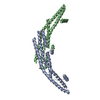 9g2uC 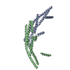 9g2wC M: map data used to model this data C: citing same article ( |
|---|---|
| Similar structure data |
-Links
 PDBj
PDBj

- Assembly
| Deposited unit | 
|
|---|---|
| 1 |
|
-Components
| #1: Protein | Mass: 40843.246 Da / Num. of mol.: 12 Source method: isolated from a genetically manipulated source Source: (gene. exp.) Homo sapiens (human) / Gene: SH3GLB1, KIAA0491, CGI-61 / Production host:  Has protein modification | N | |
|---|
-Experimental details
-Experiment
| Experiment | Method: ELECTRON MICROSCOPY |
|---|---|
| EM experiment | Aggregation state: PARTICLE / 3D reconstruction method: single particle reconstruction |
- Sample preparation
| Component | Name: Endophilin B1 dimers bound to a nanodisc / Type: COMPLEX / Entity ID: all / Source: RECOMBINANT |
|---|---|
| Molecular weight | Experimental value: NO |
| Source (natural) | Organism: Homo sapiens (human) |
| Source (recombinant) | Organism: |
| Buffer solution | pH: 7.4 / Details: 20 mM Tris-HCl, 100 mM NaCl, 0.5 mM EDTA, pH 7.4 |
| Specimen | Conc.: 1.8 mg/ml / Embedding applied: NO / Shadowing applied: NO / Staining applied: NO / Vitrification applied: YES |
| Specimen support | Grid material: COPPER / Grid mesh size: 200 divisions/in. / Grid type: Quantifoil R1.2/1.3 |
| Vitrification | Instrument: FEI VITROBOT MARK IV / Cryogen name: ETHANE / Humidity: 95 % / Chamber temperature: 277 K |
- Electron microscopy imaging
| Experimental equipment |  Model: Titan Krios / Image courtesy: FEI Company |
|---|---|
| Microscopy | Model: FEI TITAN KRIOS |
| Electron gun | Electron source:  FIELD EMISSION GUN / Accelerating voltage: 300 kV / Illumination mode: FLOOD BEAM FIELD EMISSION GUN / Accelerating voltage: 300 kV / Illumination mode: FLOOD BEAM |
| Electron lens | Mode: BRIGHT FIELD / Nominal magnification: 130000 X / Nominal defocus max: 2200 nm / Nominal defocus min: 1200 nm / Cs: 2.7 mm |
| Specimen holder | Cryogen: NITROGEN |
| Image recording | Electron dose: 40 e/Å2 / Film or detector model: GATAN K3 BIOQUANTUM (6k x 4k) |
| EM imaging optics | Energyfilter name: GIF Bioquantum / Energyfilter slit width: 20 eV |
- Processing
| EM software |
| ||||||||||||||||||||||||
|---|---|---|---|---|---|---|---|---|---|---|---|---|---|---|---|---|---|---|---|---|---|---|---|---|---|
| CTF correction | Type: PHASE FLIPPING AND AMPLITUDE CORRECTION | ||||||||||||||||||||||||
| Particle selection | Num. of particles selected: 5026978 | ||||||||||||||||||||||||
| Symmetry | Point symmetry: C1 (asymmetric) | ||||||||||||||||||||||||
| 3D reconstruction | Resolution: 3.88 Å / Resolution method: FSC 0.143 CUT-OFF / Num. of particles: 273120 / Algorithm: FOURIER SPACE / Symmetry type: POINT | ||||||||||||||||||||||||
| Atomic model building | Protocol: RIGID BODY FIT / Space: REAL Details: Experimental models determined from locally refined maps were rigid body fitted into the consensus map. | ||||||||||||||||||||||||
| Atomic model building | Accession code: AF-Q9Y371-F1-model_v4 / Chain residue range: 11-252 / Source name: AlphaFold / Type: in silico model |
