H2020 Marie Curie Actions of the European Commission
PAN.BFB.S.BDN.315.022.2022
European Union
Citation
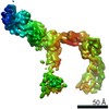
Journal: Open Biol / Year: 2025 Title: CryoEM structure and small-angle X-ray scattering analyses of porcine retinol-binding protein 3. Authors: Vineeta Kaushik / Luca Gessa / Nelam Kumar / Matyáš Pinkas / Mariusz Czarnocki-Cieciura / Krzysztof Palczewski / Jiří Nováček / Humberto Fernandes / Abstract: The vertebrate visual cycle hinges on enzymatically converting all--retinol (at-ROL) into 11--retinal (11c-RAL), the chromophore that binds to opsins in photoreceptors, forming light-responsive ...The vertebrate visual cycle hinges on enzymatically converting all--retinol (at-ROL) into 11--retinal (11c-RAL), the chromophore that binds to opsins in photoreceptors, forming light-responsive pigments. When struck by a photon, these pigments activate the phototransduction pathway and initiate the process of vision. The enzymatic isomerization of at-ROL, crucial for restoring the visual pigments and preparing them to receive new light stimuli, relies on various enzymes found in both the photoreceptors and retinal pigment epithelium cells. To function effectively, retinoids must shuttle between these two cell types. Retinol-binding protein 3 (RBP3), located in the interphotoreceptor matrix, probably plays a pivotal role in this transport mechanism. Comprised of four retinoid-binding modules, RBP3 also binds fatty acids, potentially aiding retinal function by facilitating the loading and unloading of different retinoids at specific cell types thereby directing the cycle. In this study, we present a 3.67 Å cryoEM structure of porcine RBP3, along with molecular docking analysis and corroborative in-solution small-angle X-ray scattering data for titration of RBP3 with relevant ligands, that also give insights on RBP3 conformational adaptability.
History
Deposition
Jun 19, 2024
Deposition site: PDBE / Processing site: PDBE
Revision 1.0
Feb 5, 2025
Provider: repository / Type: Initial release
Revision 1.1
Jul 2, 2025
Group: Data collection / Category: em_software / Item: _em_software.name
Mass: 138621.625 Da / Num. of mol.: 1 Source method: isolated from a genetically manipulated source Source: (gene. exp.) Sus scrofa (pig) / Gene: RBP3 / Production host: Sus scrofa (pig) / References: UniProt: A0A287A908
Has protein modification
N
-
Experimental details
-
Experiment
Experiment
Method: ELECTRON MICROSCOPY
EM experiment
Aggregation state: PARTICLE / 3D reconstruction method: single particle reconstruction
-
Sample preparation
Component
Name: Retinol binding protein 3 / Type: ORGANELLE OR CELLULAR COMPONENT / Details: Retinol binding protein 3 / Entity ID: all / Source: NATURAL
Molecular weight
Value: 138.487 kDa/nm / Experimental value: NO
Source (natural)
Organism: Sus scrofa (Pig)
Buffer solution
pH: 8
Buffer component
ID
Conc.
Name
Buffer-ID
1
50mM
HEPES
1
2
300mM
NCl
1
3
1mM
DTT
1
4
0.01 %
DDM
1
5
0.5 %
CHAPSO
1
Specimen
Conc.: 0.8 mg/ml / Embedding applied: NO / Shadowing applied: NO / Staining applied: NO / Vitrification applied: YES
Average exposure time: 2 sec. / Electron dose: 40 e/Å2 / Film or detector model: GATAN K3 BIOCONTINUUM (6k x 4k) / Num. of grids imaged: 2 / Num. of real images: 23606
EM imaging optics
Energyfilter name: GIF Bioquantum / Energyfilter slit width: 10 eV
Image scans
Width: 5760 / Height: 4092
-
Processing
EM software
ID
Name
Category
Details (eV)
1
cryoSPARC
particleselection
templatepicker
2
SerialEM
imageacquisition
4
CTFFIND
CTFcorrection
8
PHENIX
modelrefinement
12
cryoSPARC
classification
HeteroRefinement
13
cryoSPARC
3Dreconstruction
LocalRefinement
CTF correction
Type: PHASE FLIPPING AND AMPLITUDE CORRECTION
Particle selection
Num. of particles selected: 7462000
Symmetry
Point symmetry: C1 (asymmetric)
3D reconstruction
Resolution: 3.67 Å / Resolution method: FSC 0.143 CUT-OFF / Num. of particles: 611246 / Algorithm: FOURIER SPACE / Symmetry type: POINT
Refine LS restraints
Refine-ID
Type
Dev ideal
Number
ELECTRONMICROSCOPY
f_bond_d
0.002
8492
ELECTRONMICROSCOPY
f_angle_d
0.51
11567
ELECTRONMICROSCOPY
f_dihedral_angle_d
3.778
1167
ELECTRONMICROSCOPY
f_chiral_restr
0.041
1371
ELECTRONMICROSCOPY
f_plane_restr
0.003
1482
+
About Yorodumi
-
News
-
Feb 9, 2022. New format data for meta-information of EMDB entries
New format data for meta-information of EMDB entries
Version 3 of the EMDB header file is now the official format.
The previous official version 1.9 will be removed from the archive.
In the structure databanks used in Yorodumi, some data are registered as the other names, "COVID-19 virus" and "2019-nCoV". Here are the details of the virus and the list of structure data.
Jan 31, 2019. EMDB accession codes are about to change! (news from PDBe EMDB page)
EMDB accession codes are about to change! (news from PDBe EMDB page)
The allocation of 4 digits for EMDB accession codes will soon come to an end. Whilst these codes will remain in use, new EMDB accession codes will include an additional digit and will expand incrementally as the available range of codes is exhausted. The current 4-digit format prefixed with “EMD-” (i.e. EMD-XXXX) will advance to a 5-digit format (i.e. EMD-XXXXX), and so on. It is currently estimated that the 4-digit codes will be depleted around Spring 2019, at which point the 5-digit format will come into force.
The EM Navigator/Yorodumi systems omit the EMD- prefix.
Related info.:Q: What is EMD? / ID/Accession-code notation in Yorodumi/EM Navigator
Yorodumi is a browser for structure data from EMDB, PDB, SASBDB, etc.
This page is also the successor to EM Navigator detail page, and also detail information page/front-end page for Omokage search.
The word "yorodu" (or yorozu) is an old Japanese word meaning "ten thousand". "mi" (miru) is to see.
Related info.:EMDB / PDB / SASBDB / Comparison of 3 databanks / Yorodumi Search / Aug 31, 2016. New EM Navigator & Yorodumi / Yorodumi Papers / Jmol/JSmol / Function and homology information / Changes in new EM Navigator and Yorodumi
 Movie
Movie Controller


 Open data
 Basic information
 Components
 Keywords
 Function and homology information
 Authors
 Poland, European Union, 3items
  Citation

 Structure visualization
 Downloads & links
 Other downloads
 PDBj
PDBj Assembly
 Sample preparation
 Electron microscopy imaging
 FIELD EMISSION GUN / Accelerating voltage: 300 kV / Illumination mode: FLOOD BEAM
 Processing