 Movie
Movie Controller

[English] 日本語
 Yorodumi
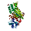
- PDB-9fqn: Crystal structure of phosphoserine phosphatase (SerB) from Brucel... -
+ Open data

- Basic information
| Entry | Database: PDB / ID: 9fqn | ||||||
|---|---|---|---|---|---|---|---|
| Title | Crystal structure of phosphoserine phosphatase (SerB) from Brucella melitensis in complex with L-Ser and Magnesium | ||||||
 Components Components | Phosphoserine phosphatase | ||||||
 Keywords Keywords | HYDROLASE / Complex / Serine biosynthesis / Magnesium cofactor / L-Serine / Product | ||||||
| Function / homology |  Function and homology information Function and homology informationphosphoserine phosphatase / L-phosphoserine phosphatase activity / L-serine biosynthetic process / magnesium ion binding / cytoplasm Similarity search - Function | ||||||
| Biological species |  Brucella melitensis (bacteria) Brucella melitensis (bacteria) | ||||||
| Method |  X-RAY DIFFRACTION / SYNCHROTRON / MOLECULAR REPLACEMENT / Resolution: 2.11 Å X-RAY DIFFRACTION / SYNCHROTRON / MOLECULAR REPLACEMENT / Resolution: 2.11 Å | ||||||
 Authors Authors | Scaillet, T. / Wouters, J. | ||||||
| Funding support | 1items
| ||||||
 Citation Citation | Journal: Proteins / Year: 2026 Title: Structural and Enzymological Characterization of Phosphoserine Phosphatase From Brucella melitensis. Authors: Scaillet, T. / Pierson, E. / Fillet, M. / Wouters, J. | ||||||
| History |
|
- Structure visualization
| Structure viewer | Molecule: MolmilJmol/JSmol |
|---|
- Downloads & links
-Download
| PDBx/mmCIF format | 9fqn.cif.gz | 92.9 KB | Display | PDBx/mmCIF format |
|---|---|---|---|---|
| PDB format | pdb9fqn.ent.gz | 56.1 KB | Display | PDB format |
| PDBx/mmJSON format | 9fqn.json.gz | Tree view | PDBx/mmJSON format | |
| Others |  Other downloads Other downloads |
-Validation report
| Arichive directory | https://data.pdbj.org/pub/pdb/validation_reports/fq/9fqnftp://data.pdbj.org/pub/pdb/validation_reports/fq/9fqn | HTTPS FTP |
|---|
-Related structure data


-Links
 PDBj
PDBj- Assembly
| Deposited unit | 
| ||||||||||||
|---|---|---|---|---|---|---|---|---|---|---|---|---|---|
| 1 |
| ||||||||||||
| Unit cell |
| ||||||||||||
| Components on special symmetry positions |
|
-Components
-Protein , 1 types, 1 molecules A
| #1: Protein | Mass: 32549.479 Da / Num. of mol.: 1 Source method: isolated from a genetically manipulated source Source: (gene. exp.) Brucella melitensis (bacteria) / Gene: BMEI0615 / Production host: |
|---|
-Non-polymers , 6 types, 209 molecules 


| #2: Chemical | ChemComp-SER /  Type: L-peptide linking / Mass: 105.093 Da / Num. of mol.: 1 / Source method: obtained synthetically / Formula: C3H7NO3 / Feature type: SUBJECT OF INVESTIGATION Type: L-peptide linking / Mass: 105.093 Da / Num. of mol.: 1 / Source method: obtained synthetically / Formula: C3H7NO3 / Feature type: SUBJECT OF INVESTIGATION | ||||||||
|---|---|---|---|---|---|---|---|---|---|
| #3: Chemical |  Mass: 106.120 Da / Num. of mol.: 2 / Source method: obtained synthetically / Formula: C4H10O3 Mass: 106.120 Da / Num. of mol.: 2 / Source method: obtained synthetically / Formula: C4H10O3#4: Chemical | ChemComp-GOL / |  Mass: 92.094 Da / Num. of mol.: 1 / Source method: obtained synthetically / Formula: C3H8O3 Mass: 92.094 Da / Num. of mol.: 1 / Source method: obtained synthetically / Formula: C3H8O3#5: Chemical | ChemComp-MG / |  Mass: 24.305 Da / Num. of mol.: 1 / Source method: obtained synthetically / Formula: Mg Mass: 24.305 Da / Num. of mol.: 1 / Source method: obtained synthetically / Formula: Mg#6: Chemical |  Mass: 35.453 Da / Num. of mol.: 2 / Source method: obtained synthetically / Formula: Cl Mass: 35.453 Da / Num. of mol.: 2 / Source method: obtained synthetically / Formula: Cl#7: Water | ChemComp-HOH / | Mass: 18.015 Da / Num. of mol.: 202 / Source method: isolated from a natural source / Formula: H2O |
-Details
| Has ligand of interest | Y |
|---|---|
| Has protein modification | N |
-Experimental details
-Experiment
| Experiment | Method: X-RAY DIFFRACTION / Number of used crystals: 1 |
|---|
- Sample preparation
| Crystal | Density Matthews: 3.54 Å3/Da / Density % sol: 65.3 % |
|---|---|
| Crystal grow | Temperature: 293 K / Method: vapor diffusion / pH: 8 Details: 0.2M sodium malonate, 0.1M Bis-tris propane, PEG3350 PH range: 6.5 - 8.0 / Temp details: Room temperature |
-Data collection
| Diffraction | Mean temperature: 100 K / Serial crystal experiment: N |
|---|---|
| Diffraction source | Source: SYNCHROTRON / Site: SOLEIL  / Beamline: PROXIMA 2 / Wavelength: 1.0439 Å / Beamline: PROXIMA 2 / Wavelength: 1.0439 Å |
| Detector | Type: DECTRIS EIGER X 9M / Detector: PIXEL / Date: Mar 30, 2023 |
| Radiation | Protocol: SINGLE WAVELENGTH / Monochromatic (M) / Laue (L): M / Scattering type: x-ray |
| Radiation wavelength | Wavelength: 1.0439 Å / Relative weight: 1 |
| Reflection | Resolution: 2.11→45.17 Å / Num. obs: 27894 / % possible obs: 99.84 % / Redundancy: 40.1 % / Biso Wilson estimate: 48.65 Å2 / CC1/2: 1 / CC star: 1 / Rmerge(I) obs: 0.08128 / Rpim(I) all: 0.01306 / Rrim(I) all: 0.08233 / Net I/σ(I): 35.36 |
| Reflection shell | Resolution: 2.11→2.19 Å / Redundancy: 38.3 % / Rmerge(I) obs: 1.374 / Mean I/σ(I) obs: 2.78 / Num. unique obs: 2749 / CC1/2: 0.856 / CC star: 0.96 / Rpim(I) all: 0.2232 / Rrim(I) all: 1.393 / % possible all: 98.6 |
- Processing
| Software |
| |||||||||||||||||||||||||||||||||||||||||||||||||||||||||||||||||||||||||||||
|---|---|---|---|---|---|---|---|---|---|---|---|---|---|---|---|---|---|---|---|---|---|---|---|---|---|---|---|---|---|---|---|---|---|---|---|---|---|---|---|---|---|---|---|---|---|---|---|---|---|---|---|---|---|---|---|---|---|---|---|---|---|---|---|---|---|---|---|---|---|---|---|---|---|---|---|---|---|---|
| Refinement | Method to determine structure: MOLECULAR REPLACEMENT / Resolution: 2.11→45.17 Å / SU ML: 0.3229 / Cross valid method: FREE R-VALUE / σ(F): 1.38 / Phase error: 28.8999 Stereochemistry target values: GeoStd + Monomer Library + CDL v1.2
| |||||||||||||||||||||||||||||||||||||||||||||||||||||||||||||||||||||||||||||
| Solvent computation | Shrinkage radii: 0.9 Å / VDW probe radii: 1.1 Å / Solvent model: FLAT BULK SOLVENT MODEL | |||||||||||||||||||||||||||||||||||||||||||||||||||||||||||||||||||||||||||||
| Displacement parameters | Biso mean: 53.17 Å2 | |||||||||||||||||||||||||||||||||||||||||||||||||||||||||||||||||||||||||||||
| Refinement step | Cycle: LAST / Resolution: 2.11→45.17 Å
| |||||||||||||||||||||||||||||||||||||||||||||||||||||||||||||||||||||||||||||
| Refine LS restraints |
| |||||||||||||||||||||||||||||||||||||||||||||||||||||||||||||||||||||||||||||
| LS refinement shell |
|
