 Movie
Movie Controller


+ Open data

- Basic information
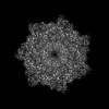
| Entry | Database: PDB / ID: 9fmx | ||||||||||||
|---|---|---|---|---|---|---|---|---|---|---|---|---|---|
| Title | Aerolysin Y221G - prepore | ||||||||||||
 Components Components | Aerolysin | ||||||||||||
 Keywords Keywords | TOXIN / pore forming toxin / aerolysin / cryo-EM | ||||||||||||
| Function / homology |  Function and homology information Function and homology informationsymbiont-mediated cytolysis of host cell / toxin activity / host cell plasma membrane / extracellular region / identical protein binding / membrane Similarity search - Function | ||||||||||||
| Biological species |  Aeromonas hydrophila (bacteria) Aeromonas hydrophila (bacteria) | ||||||||||||
| Method | ELECTRON MICROSCOPY / single particle reconstruction / cryo EM / Resolution: 2.2 Å | ||||||||||||
 Authors Authors | Iacovache, I. / Zuber, B. | ||||||||||||
| Funding support |  Switzerland, 3items Switzerland, 3items
| ||||||||||||
 Citation Citation | Journal: J Am Chem Soc / Year: 2025 Title: Aerolysin Nanopore Structures Revealed at High Resolution in a Lipid Environment. Authors: Jana S Anton / Ioan Iacovache / Juan F Bada Juarez / Luciano A Abriata / Louis W Perrin / Chan Cao / Maria J Marcaida / Benoît Zuber / Matteo Dal Peraro / Abstract: Aerolysin is a β-pore-forming toxin produced by most Aeromonas bacteria, which has attracted large attention in the field of nanopore sensing due to its narrow and charged pore lumen. Structurally ...Aerolysin is a β-pore-forming toxin produced by most Aeromonas bacteria, which has attracted large attention in the field of nanopore sensing due to its narrow and charged pore lumen. Structurally similar proteins, belonging to the aerolysin-like family, are present throughout all kingdoms of life, but very few of them have been structurally characterized in a lipid environment. Here, we present the first high-resolution atomic cryo-EM structures of aerolysin prepore and pore in a membrane-like environment. These structures allow the identification of key interactions, which are relevant for understanding the pore formation mechanism and for correctly positioning the pore β-barrel and its anchoring β-turn motif in the membrane. Moreover, we elucidate at high resolution the architecture of key pore mutations and precisely identify four constriction rings in the pore lumen that are highly relevant for nanopore sensing experiments. #1: Journal: Acta Crystallogr D Struct Biol / Year: 2019 Title: Macromolecular structure determination using X-rays, neutrons and electrons: recent developments in Phenix. Authors: Dorothee Liebschner / Pavel V Afonine / Matthew L Baker / Gábor Bunkóczi / Vincent B Chen / Tristan I Croll / Bradley Hintze / Li Wei Hung / Swati Jain / Airlie J McCoy / Nigel W Moriarty ...Authors: Dorothee Liebschner / Pavel V Afonine / Matthew L Baker / Gábor Bunkóczi / Vincent B Chen / Tristan I Croll / Bradley Hintze / Li Wei Hung / Swati Jain / Airlie J McCoy / Nigel W Moriarty / Robert D Oeffner / Billy K Poon / Michael G Prisant / Randy J Read / Jane S Richardson / David C Richardson / Massimo D Sammito / Oleg V Sobolev / Duncan H Stockwell / Thomas C Terwilliger / Alexandre G Urzhumtsev / Lizbeth L Videau / Christopher J Williams / Paul D Adams /    Abstract: Diffraction (X-ray, neutron and electron) and electron cryo-microscopy are powerful methods to determine three-dimensional macromolecular structures, which are required to understand biological ...Diffraction (X-ray, neutron and electron) and electron cryo-microscopy are powerful methods to determine three-dimensional macromolecular structures, which are required to understand biological processes and to develop new therapeutics against diseases. The overall structure-solution workflow is similar for these techniques, but nuances exist because the properties of the reduced experimental data are different. Software tools for structure determination should therefore be tailored for each method. Phenix is a comprehensive software package for macromolecular structure determination that handles data from any of these techniques. Tasks performed with Phenix include data-quality assessment, map improvement, model building, the validation/rebuilding/refinement cycle and deposition. Each tool caters to the type of experimental data. The design of Phenix emphasizes the automation of procedures, where possible, to minimize repetitive and time-consuming manual tasks, while default parameters are chosen to encourage best practice. A graphical user interface provides access to many command-line features of Phenix and streamlines the transition between programs, project tracking and re-running of previous tasks. | ||||||||||||
| History |
|
- Structure visualization
| Structure viewer | Molecule: MolmilJmol/JSmol |
|---|
- Downloads & links
-Download
| PDBx/mmCIF format | 9fmx.cif.gz | 2 MB | Display | PDBx/mmCIF format |
|---|---|---|---|---|
| PDB format | pdb9fmx.ent.gz | Display | PDB format | |
| PDBx/mmJSON format | 9fmx.json.gz | Tree view | PDBx/mmJSON format | |
| Others |  Other downloads Other downloads |
-Validation report
| Summary document | 9fmx_validation.pdf.gz | 1.3 MB | Display | wwPDB validaton report |
|---|---|---|---|---|
| Full document | 9fmx_full_validation.pdf.gz | 1.3 MB | Display | |
| Data in XML | 9fmx_validation.xml.gz | 148 KB | Display | |
| Data in CIF | 9fmx_validation.cif.gz | 227 KB | Display | |
| Arichive directory | https://data.pdbj.org/pub/pdb/validation_reports/fm/9fmxftp://data.pdbj.org/pub/pdb/validation_reports/fm/9fmx | HTTPS FTP |
-Related structure data
| Related structure data |  50576MC  9fm6C  9fmlC  9fnpC  9fnqC M: map data used to model this data C: citing same article ( |
|---|---|
| Similar structure data |


-Links
 PDBj
PDBj


- Assembly
| Deposited unit | 
|
|---|---|
| 1 |
|
-Components
| #1: Protein | Mass: 52945.473 Da / Num. of mol.: 14 / Mutation: Y221G Source method: isolated from a genetically manipulated source Source: (gene. exp.) Aeromonas hydrophila (bacteria) / Gene: aerA / Production host: Has protein modification | Y | |
|---|
-Experimental details
-Experiment
| Experiment | Method: ELECTRON MICROSCOPY |
|---|---|
| EM experiment | Aggregation state: PARTICLE / 3D reconstruction method: single particle reconstruction |
- Sample preparation
| Component | Name: aerolysin pre-pore Y221G double heptamer / Type: COMPLEX Details: Heptamerization was induced by trypsinization of proaerolysin Y221G. Entity ID: all / Source: RECOMBINANT |
|---|---|
| Molecular weight | Value: 0.7 MDa / Experimental value: NO |
| Source (natural) | Organism: Aeromonas hydrophila (bacteria) |
| Source (recombinant) | Organism: |
| Buffer solution | pH: 7.4 / Details: 20mM Hepes 7.4 100mM NaCl 0.05% A8-35 |
| Specimen | Conc.: 1 mg/ml / Embedding applied: NO / Shadowing applied: NO / Staining applied: NO / Vitrification applied: YES |
| Specimen support | Grid type: Quantifoil |
| Vitrification | Instrument: FEI VITROBOT MARK IV / Cryogen name: ETHANE / Humidity: 100 % / Chamber temperature: 298 K |
- Electron microscopy imaging
| Experimental equipment |  Model: Titan Krios / Image courtesy: FEI Company |
|---|---|
| Microscopy | Model: FEI TITAN KRIOS |
| Electron gun | Electron source:  FIELD EMISSION GUN / Accelerating voltage: 300 kV / Illumination mode: FLOOD BEAM FIELD EMISSION GUN / Accelerating voltage: 300 kV / Illumination mode: FLOOD BEAM |
| Electron lens | Mode: BRIGHT FIELD / Nominal defocus max: 3000 nm / Nominal defocus min: 1200 nm / Cs: 2.7 mm / Alignment procedure: BASIC |
| Specimen holder | Cryogen: NITROGEN / Specimen holder model: FEI TITAN KRIOS AUTOGRID HOLDER |
| Image recording | Electron dose: 40 e/Å2 / Film or detector model: FEI FALCON IV (4k x 4k) |
| EM imaging optics | Energyfilter name: TFS Selectris / Energyfilter slit width: 20 eV |
- Processing
| EM software |
| ||||||||||||||||||||||||||||||||||||||||
|---|---|---|---|---|---|---|---|---|---|---|---|---|---|---|---|---|---|---|---|---|---|---|---|---|---|---|---|---|---|---|---|---|---|---|---|---|---|---|---|---|---|
| CTF correction | Type: PHASE FLIPPING AND AMPLITUDE CORRECTION | ||||||||||||||||||||||||||||||||||||||||
| Symmetry | Point symmetry: D7 (2x7 fold dihedral) | ||||||||||||||||||||||||||||||||||||||||
| 3D reconstruction | Resolution: 2.2 Å / Resolution method: FSC 0.143 CUT-OFF / Num. of particles: 329000 / Symmetry type: POINT | ||||||||||||||||||||||||||||||||||||||||
| Atomic model building | Protocol: OTHER Details: ModelAngelo was used with the final map to generate the first model. | ||||||||||||||||||||||||||||||||||||||||
| Atomic model building | Details: modelangelo / Source name: Other / Type: other | ||||||||||||||||||||||||||||||||||||||||
| Refine LS restraints |
|
