 Movie
Movie Controller


+ Open data

- Basic information
| Entry | Database: PDB / ID: 9fd6 | ||||||
|---|---|---|---|---|---|---|---|
| Title | flavin reductase ThdF in complex with NAD and FAD | ||||||
 Components Components | Flavin reductase | ||||||
 Keywords Keywords | FLAVOPROTEIN / flavin reductase / FAD / oxidoreductase / NADH:FAD oxdioreductase | ||||||
| Function / homology |  Function and homology information Function and homology information | ||||||
| Biological species |  Streptomyces albogriseolus (bacteria) Streptomyces albogriseolus (bacteria) | ||||||
| Method |  X-RAY DIFFRACTION / SYNCHROTRON / FOURIER SYNTHESIS / Resolution: 1.43 Å X-RAY DIFFRACTION / SYNCHROTRON / FOURIER SYNTHESIS / Resolution: 1.43 Å | ||||||
 Authors Authors | Bork, S. / Nagel, M.F. / Keller, W. / Niemann, H.H. | ||||||
| Funding support |  Germany, 1items Germany, 1items
| ||||||
 Citation Citation | Journal: J.Biol.Chem. / Year: 2024 Title: The NADH-dependent flavin reductase ThdF follows an ordered sequential mechanism though crystal structures reveal two FAD molecules in the active site. Authors: Horstmeier, H.J. / Bork, S. / Nagel, M.F. / Keller, W. / Spross, J. / Diepold, N. / Ruppel, M. / Kottke, T. / Niemann, H.H. | ||||||
| History |
|
- Structure visualization
| Structure viewer | Molecule: MolmilJmol/JSmol |
|---|
- Downloads & links
-Download
| PDBx/mmCIF format | 9fd6.cif.gz | 430 KB | Display | PDBx/mmCIF format |
|---|---|---|---|---|
| PDB format | pdb9fd6.ent.gz | 366.2 KB | Display | PDB format |
| PDBx/mmJSON format | 9fd6.json.gz | Tree view | PDBx/mmJSON format | |
| Others |  Other downloads Other downloads |
-Validation report
| Summary document | 9fd6_validation.pdf.gz | 5.4 MB | Display | wwPDB validaton report |
|---|---|---|---|---|
| Full document | 9fd6_full_validation.pdf.gz | 5.4 MB | Display | |
| Data in XML | 9fd6_validation.xml.gz | 40.8 KB | Display | |
| Data in CIF | 9fd6_validation.cif.gz | 54.9 KB | Display | |
| Arichive directory | https://data.pdbj.org/pub/pdb/validation_reports/fd/9fd6ftp://data.pdbj.org/pub/pdb/validation_reports/fd/9fd6 | HTTPS FTP |
-Related structure data
| Related structure data |  9fd4C 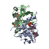 9fd5C C: citing same article ( |
|---|---|
| Similar structure data | |
| Experimental dataset #1 | Data reference: 10.15785/SBGRID/1111 / Data set type: diffraction image data |
-Links
 PDBj
PDBj

- Assembly
| Deposited unit | 
| ||||||||||
|---|---|---|---|---|---|---|---|---|---|---|---|
| 1 | 
| ||||||||||
| 2 | 
| ||||||||||
| Unit cell |
|
-Components
| #1: Protein | Mass: 20327.689 Da / Num. of mol.: 4 Source method: isolated from a genetically manipulated source Source: (gene. exp.) Streptomyces albogriseolus (bacteria) / Gene: thdF / Plasmid: pETM-11 / Production host: #2: Chemical | ChemComp-FAD /   Mass: 785.550 Da / Num. of mol.: 4 / Source method: obtained synthetically / Formula: C27H33N9O15P2 / Feature type: SUBJECT OF INVESTIGATION / Comment: FAD*YM Mass: 785.550 Da / Num. of mol.: 4 / Source method: obtained synthetically / Formula: C27H33N9O15P2 / Feature type: SUBJECT OF INVESTIGATION / Comment: FAD*YM#3: Chemical | ChemComp-NAI /   Mass: 665.441 Da / Num. of mol.: 4 / Source method: obtained synthetically / Formula: C21H29N7O14P2 / Feature type: SUBJECT OF INVESTIGATION Mass: 665.441 Da / Num. of mol.: 4 / Source method: obtained synthetically / Formula: C21H29N7O14P2 / Feature type: SUBJECT OF INVESTIGATION#4: Water | ChemComp-HOH / |  Mass: 18.015 Da / Num. of mol.: 659 / Source method: isolated from a natural source / Formula: H2O Mass: 18.015 Da / Num. of mol.: 659 / Source method: isolated from a natural source / Formula: H2OHas ligand of interest | Y | Has protein modification | N | |
|---|
-Experimental details
-Experiment
| Experiment | Method: X-RAY DIFFRACTION / Number of used crystals: 1 |
|---|
- Sample preparation
| Crystal | Density Matthews: 1.95 Å3/Da / Density % sol: 36.92 % |
|---|---|
| Crystal grow | Temperature: 293 K / Method: vapor diffusion, sitting drop Details: D12 JCSG++ (Jena Bioscience); 40 mM KH2PO4, 20 % (v/v) glycerol, 16 % (w/v) PEG 8000; 18 mg/mL ThdF (50 mM HEPES pH 7.0, 20 mM NaCl, 1 mM DTT), 1:1800 (mass ratio) elastase added; 600 nL ...Details: D12 JCSG++ (Jena Bioscience); 40 mM KH2PO4, 20 % (v/v) glycerol, 16 % (w/v) PEG 8000; 18 mg/mL ThdF (50 mM HEPES pH 7.0, 20 mM NaCl, 1 mM DTT), 1:1800 (mass ratio) elastase added; 600 nL protein + 300 nL reservoir; soaking with 250 nL 100 mM NADH and 250 nL 500 mM dithionite |
-Data collection
| Diffraction | Mean temperature: 100 K / Serial crystal experiment: N |
|---|---|
| Diffraction source | Source: SYNCHROTRON / Site: PETRA III, EMBL c/o DESY / Beamline: P13 (MX1) / Wavelength: 0.9762 Å |
| Detector | Type: DECTRIS EIGER X 16M / Detector: PIXEL / Date: May 13, 2023 |
| Radiation | Protocol: SINGLE WAVELENGTH / Monochromatic (M) / Laue (L): M / Scattering type: x-ray |
| Radiation wavelength | Wavelength: 0.9762 Å / Relative weight: 1 |
| Reflection | Resolution: 1.43→73.92 Å / Num. obs: 95935 / % possible obs: 80.3 % / Redundancy: 13.5 % / Biso Wilson estimate: 18.12 Å2 / CC1/2: 0.999 / Rmerge(I) obs: 0.095 / Rpim(I) all: 0.027 / Rrim(I) all: 0.098 / Net I/σ(I): 14.9 |
| Reflection shell | Resolution: 1.43→1.55 Å / Redundancy: 13.8 % / Rmerge(I) obs: 1.901 / Mean I/σ(I) obs: 1.5 / Num. unique obs: 4797 / CC1/2: 0.594 / Rpim(I) all: 0.529 / Rrim(I) all: 1.974 / % possible all: 18.6 |
- Processing
| Software |
| |||||||||||||||||||||||||||||||||||||||||||||||||||||||||||||||||||||||||||||||||||||||||||||||||||||||||||||||||||||||||||||||||||||||||||||||||||||||||||||||||||||||||||||||||||||||||||||||||||||||||||||||||||||||||
|---|---|---|---|---|---|---|---|---|---|---|---|---|---|---|---|---|---|---|---|---|---|---|---|---|---|---|---|---|---|---|---|---|---|---|---|---|---|---|---|---|---|---|---|---|---|---|---|---|---|---|---|---|---|---|---|---|---|---|---|---|---|---|---|---|---|---|---|---|---|---|---|---|---|---|---|---|---|---|---|---|---|---|---|---|---|---|---|---|---|---|---|---|---|---|---|---|---|---|---|---|---|---|---|---|---|---|---|---|---|---|---|---|---|---|---|---|---|---|---|---|---|---|---|---|---|---|---|---|---|---|---|---|---|---|---|---|---|---|---|---|---|---|---|---|---|---|---|---|---|---|---|---|---|---|---|---|---|---|---|---|---|---|---|---|---|---|---|---|---|---|---|---|---|---|---|---|---|---|---|---|---|---|---|---|---|---|---|---|---|---|---|---|---|---|---|---|---|---|---|---|---|---|---|---|---|---|---|---|---|---|---|---|---|---|---|---|---|---|
| Refinement | Method to determine structure: FOURIER SYNTHESIS / Resolution: 1.43→45.74 Å / SU ML: 0.15 / Cross valid method: FREE R-VALUE / σ(F): 1.34 / Phase error: 20.86 / Stereochemistry target values: ML
| |||||||||||||||||||||||||||||||||||||||||||||||||||||||||||||||||||||||||||||||||||||||||||||||||||||||||||||||||||||||||||||||||||||||||||||||||||||||||||||||||||||||||||||||||||||||||||||||||||||||||||||||||||||||||
| Solvent computation | Shrinkage radii: 0.9 Å / VDW probe radii: 1.1 Å / Solvent model: FLAT BULK SOLVENT MODEL | |||||||||||||||||||||||||||||||||||||||||||||||||||||||||||||||||||||||||||||||||||||||||||||||||||||||||||||||||||||||||||||||||||||||||||||||||||||||||||||||||||||||||||||||||||||||||||||||||||||||||||||||||||||||||
| Refinement step | Cycle: LAST / Resolution: 1.43→45.74 Å
| |||||||||||||||||||||||||||||||||||||||||||||||||||||||||||||||||||||||||||||||||||||||||||||||||||||||||||||||||||||||||||||||||||||||||||||||||||||||||||||||||||||||||||||||||||||||||||||||||||||||||||||||||||||||||
| Refine LS restraints |
| |||||||||||||||||||||||||||||||||||||||||||||||||||||||||||||||||||||||||||||||||||||||||||||||||||||||||||||||||||||||||||||||||||||||||||||||||||||||||||||||||||||||||||||||||||||||||||||||||||||||||||||||||||||||||
| LS refinement shell |
|
