Movie
Movie Controller


+ Open data

- Basic information
| Entry | Database: PDB / ID: 9f9y | |||||||||
|---|---|---|---|---|---|---|---|---|---|---|
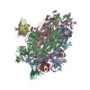
| Title | SARS-CoV-2 BA-2.87.1 Spike ectodomain | |||||||||
 Components Components | Spike glycoprotein,Fibritin | |||||||||
 Keywords Keywords | VIRAL PROTEIN / immune system / SARS-CoV-2 / RBD / Spike / glycoprotein / BA.2.87.1 / receptor / coronavirus-2 / N-terminal domain / Supersite | |||||||||
| Function / homology |  Function and homology information Function and homology informationvirion component / symbiont-mediated disruption of host tissue / Maturation of spike protein / Translation of Structural Proteins / Virion Assembly and Release / host cell surface / host extracellular region / symbiont-mediated-mediated suppression of host tetherin activity / Induction of Cell-Cell Fusion / structural constituent of virion ...virion component / symbiont-mediated disruption of host tissue / Maturation of spike protein / Translation of Structural Proteins / Virion Assembly and Release / host cell surface / host extracellular region / symbiont-mediated-mediated suppression of host tetherin activity / Induction of Cell-Cell Fusion / structural constituent of virion / positive regulation of viral entry into host cell / membrane fusion / host cell endoplasmic reticulum-Golgi intermediate compartment membrane / Attachment and Entry / entry receptor-mediated virion attachment to host cell / receptor-mediated virion attachment to host cell / host cell surface receptor binding / symbiont-mediated suppression of host innate immune response / endocytosis involved in viral entry into host cell / receptor ligand activity / fusion of virus membrane with host plasma membrane / fusion of virus membrane with host endosome membrane / viral envelope / symbiont entry into host cell / virion attachment to host cell / host cell plasma membrane / SARS-CoV-2 activates/modulates innate and adaptive immune responses / virion membrane / membrane / identical protein binding / plasma membrane Similarity search - Function | |||||||||
| Biological species |   Severe acute respiratory syndrome coronavirus 2Enterobacteria phage T4 (virus) Severe acute respiratory syndrome coronavirus 2Enterobacteria phage T4 (virus) | |||||||||
| Method | ELECTRON MICROSCOPY / single particle reconstruction / cryo EM / Resolution: 2.8 Å | |||||||||
 Authors Authors | Ren, J. / Stuart, D.I. / Duyvesteyn, H.M.E. | |||||||||
| Funding support |  United Kingdom, 2items United Kingdom, 2items
| |||||||||
 Citation Citation | Journal: Structure / Year: 2024 Title: Concerted deletions eliminate a neutralizing supersite in SARS-CoV-2 BA.2.87.1 spike. Authors: Helen M E Duyvesteyn / Aiste Dijokaite-Guraliuc / Chang Liu / Piyada Supasa / Barbara Kronsteiner / Katie Jeffery / Lizzie Stafford / Paul Klenerman / Susanna J Dunachie / / Juthathip ...Authors: Helen M E Duyvesteyn / Aiste Dijokaite-Guraliuc / Chang Liu / Piyada Supasa / Barbara Kronsteiner / Katie Jeffery / Lizzie Stafford / Paul Klenerman / Susanna J Dunachie / / Juthathip Mongkolsapaya / Elizabeth E Fry / Jingshan Ren / David I Stuart / Gavin R Screaton /  Abstract: BA.2.87.1 represents a major shift in the BA.2 lineage of severe acute respiratory syndrome coronavirus 2 (SARS-CoV-2) and is unusual in having two lengthy deletions of polypeptide in the spike (S) ...BA.2.87.1 represents a major shift in the BA.2 lineage of severe acute respiratory syndrome coronavirus 2 (SARS-CoV-2) and is unusual in having two lengthy deletions of polypeptide in the spike (S) protein, one of which removes a beta-strand. Here we investigate its neutralization by a variety of sera from infected and vaccinated individuals and determine its spike (S) ectodomain structure. The BA.2.87.1 receptor binding domain (RBD) is structurally conserved and the RBDs are tightly packed in an "all-down" conformation with a small rotation relative to the trimer axis as compared to the closest previously observed conformation. The N-terminal domain (NTD) maintains a remarkably similar structure overall; however, the rearrangements resulting from the deletions essentially destroy the so-called supersite epitope and eliminate one glycan site, while a mutation creates an additional glycan site, effectively shielding another NTD epitope. BA.2.87.1 is relatively easily neutralized but acquisition of additional mutations in the RBD could increase antibody escape allowing it to become a dominant sub-lineage. #1: Journal: Acta Crystallogr., Sect. D: Biol. Crystallogr. / Year: 2019Title: Macromolecular structure determination using X-rays, neutrons and electrons: recent developments in Phenix Authors: Liebschner, D. / Afonine, P.V. #2: Journal: Nature Methods / Year: 2017Title: cryoSPARC: algorithms for rapid unsupervised cryo-EM structure determination Authors: Punjani, A. / Rubinstein, J.L. | |||||||||
| History |
|
- Structure visualization
| Structure viewer | Molecule: MolmilJmol/JSmol |
|---|
- Downloads & links
-Download
| PDBx/mmCIF format | 9f9y.cif.gz | 631.2 KB | Display | PDBx/mmCIF format |
|---|---|---|---|---|
| PDB format | pdb9f9y.ent.gz | 508.8 KB | Display | PDB format |
| PDBx/mmJSON format | 9f9y.json.gz | Tree view | PDBx/mmJSON format | |
| Others |  Other downloads Other downloads |
-Validation report
| Arichive directory | https://data.pdbj.org/pub/pdb/validation_reports/f9/9f9yftp://data.pdbj.org/pub/pdb/validation_reports/f9/9f9y | HTTPS FTP |
|---|
-Related structure data
| Related structure data |  50263MC M: map data used to model this data C: citing same article ( |
|---|---|
| Similar structure data |
-Links
 PDBj
PDBj


- Assembly
| Deposited unit | 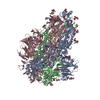
|
|---|---|
| 1 |
|
-Components
| #1: Protein | Mass: 139502.484 Da / Num. of mol.: 3 Source method: isolated from a genetically manipulated source Source: (gene. exp.) Severe acute respiratory syndrome coronavirus 2, (gene. exp.) Enterobacteria phage T4 (virus)Gene: S, 2, wac / Cell line (production host): Human Embryonic Kidney 293T / Production host:  Homo sapiens (human) / References: UniProt: P0DTC2, UniProt: P10104 Homo sapiens (human) / References: UniProt: P0DTC2, UniProt: P10104#2: Polysaccharide | 2-acetamido-2-deoxy-beta-D-glucopyranose-(1-4)-2-acetamido-2-deoxy-beta-D-glucopyranose Source method: isolated from a genetically manipulated source #3: Sugar | ChemComp-NAG /   Type: D-saccharide, beta linking / Mass: 221.208 Da / Num. of mol.: 39 / Source method: obtained synthetically / Formula: C8H15NO6 Type: D-saccharide, beta linking / Mass: 221.208 Da / Num. of mol.: 39 / Source method: obtained synthetically / Formula: C8H15NO6Has ligand of interest | N | Has protein modification | Y | |
|---|
-Experimental details
-Experiment
| Experiment | Method: ELECTRON MICROSCOPY |
|---|---|
| EM experiment | Aggregation state: PARTICLE / 3D reconstruction method: single particle reconstruction |
- Sample preparation
| Component | Name: BA-2.87.1 variant SARS-CoV2 S protein / Type: COMPLEX Details: Spike protein recombinantly expressed using sequence of human-derived BA-2.87.1 variant of SARS-CoV2. Entity ID: #1 / Source: RECOMBINANT | ||||||||||||
|---|---|---|---|---|---|---|---|---|---|---|---|---|---|
| Molecular weight | Experimental value: NO | ||||||||||||
| Source (natural) |
| ||||||||||||
| Source (recombinant) | Organism: Homo sapiens (human) | ||||||||||||
| Details of virus | Empty: YES / Enveloped: NO / Isolate: STRAIN / Type: VIRUS-LIKE PARTICLE | ||||||||||||
| Natural host | Organism: Homo sapiens | ||||||||||||
| Buffer solution | pH: 7.4 | ||||||||||||
| Specimen | Embedding applied: NO / Shadowing applied: NO / Staining applied: NO / Vitrification applied: YES / Details: BA.2.87.1 Spike | ||||||||||||
| Specimen support | Grid material: COPPER / Grid mesh size: 200 divisions/in. / Grid type: C-flat-2/1 | ||||||||||||
| Vitrification | Cryogen name: ETHANE |
- Electron microscopy imaging
| Experimental equipment |  Model: Titan Krios / Image courtesy: FEI Company |
|---|---|
| Microscopy | Model: FEI TITAN KRIOS |
| Electron gun | Electron source:  FIELD EMISSION GUN / Accelerating voltage: 300 kV / Illumination mode: OTHER FIELD EMISSION GUN / Accelerating voltage: 300 kV / Illumination mode: OTHER |
| Electron lens | Mode: BRIGHT FIELD / Nominal magnification: 165000 X / Nominal defocus max: 2600 nm / Nominal defocus min: 800 nm / Cs: 2.7 mm / C2 aperture diameter: 50 µm |
| Specimen holder | Cryogen: NITROGEN / Specimen holder model: FEI TITAN KRIOS AUTOGRID HOLDER |
| Image recording | Electron dose: 40 e/Å2 / Film or detector model: FEI FALCON IV (4k x 4k) / Num. of grids imaged: 1 / Num. of real images: 9818 / Details: Images were collected in EER format. |
| EM imaging optics | Energyfilter name: TFS Selectris X / Energyfilter slit width: 10 eV |
- Processing
| EM software |
| ||||||||||||||||||||||||||||||||||||||||||||
|---|---|---|---|---|---|---|---|---|---|---|---|---|---|---|---|---|---|---|---|---|---|---|---|---|---|---|---|---|---|---|---|---|---|---|---|---|---|---|---|---|---|---|---|---|---|
| CTF correction | Details: Determined using cryoSPARC live patch CTF correction. Type: PHASE FLIPPING AND AMPLITUDE CORRECTION | ||||||||||||||||||||||||||||||||||||||||||||
| Particle selection | Num. of particles selected: 504319 | ||||||||||||||||||||||||||||||||||||||||||||
| Symmetry | Point symmetry: C3 (3 fold cyclic) | ||||||||||||||||||||||||||||||||||||||||||||
| 3D reconstruction | Resolution: 2.8 Å / Resolution method: FSC 0.143 CUT-OFF / Num. of particles: 37441 Details: Determined in cryosparc local resolution estimation module. Num. of class averages: 1 / Symmetry type: POINT | ||||||||||||||||||||||||||||||||||||||||||||
| Atomic model building | Protocol: OTHER / Space: REAL Details: Phenix real space refinement with manual checks using coot. | ||||||||||||||||||||||||||||||||||||||||||||
| Atomic model building | PDB-ID: 8R1C Accession code: 8R1C / Source name: PDB / Type: experimental model | ||||||||||||||||||||||||||||||||||||||||||||
| Refine LS restraints |
|