 Movie
Movie Controller

[English] 日本語
 Yorodumi
- PDB-9f9h: Laser excitation effects on BR: Reprocessed Extrapolated 10ps Lig... -
+ Open data

- Basic information
| Entry | Database: PDB / ID: 9f9h | ||||||
|---|---|---|---|---|---|---|---|
| Title | Laser excitation effects on BR: Reprocessed Extrapolated 10ps Light dataset recorded at 525 GW/cm2 from Nogly et al. | ||||||
 Components Components | Bacteriorhodopsin | ||||||
 Keywords Keywords | PROTON TRANSPORT / Bacteriorhodospin / Membrane / RETINAL / TIME-RESOLVED CRYSTALLOGRAPHY / SERIAL CRYSTALLOGRAPHY / Laser excitation | ||||||
| Function / homology |  Function and homology information Function and homology informationlight-driven active monoatomic ion transmembrane transporter activity / photoreceptor activity / phototransduction / monoatomic ion channel activity / proton transmembrane transport / plasma membrane Similarity search - Function | ||||||
| Biological species |  Halobacterium salinarum (Halophile) Halobacterium salinarum (Halophile) | ||||||
| Method |  X-RAY DIFFRACTION / FREE ELECTRON LASER / MOLECULAR REPLACEMENT / Resolution: 1.66 Å X-RAY DIFFRACTION / FREE ELECTRON LASER / MOLECULAR REPLACEMENT / Resolution: 1.66 Å | ||||||
 Authors Authors | Bertrand, Q. / Weinert, T. / Standfuss, J. | ||||||
| Funding support | European Union, 1items
| ||||||
 Citation Citation | Journal: Nat Commun / Year: 2024 Title: Structural effects of high laser power densities on an early bacteriorhodopsin photocycle intermediate. Authors: Bertrand, Q. / Nogly, P. / Nango, E. / Kekilli, D. / Khusainov, G. / Furrer, A. / James, D. / Dworkowski, F. / Skopintsev, P. / Mous, S. / Martiel, I. / Borjesson, P. / Ortolani, G. / Huang, ...Authors: Bertrand, Q. / Nogly, P. / Nango, E. / Kekilli, D. / Khusainov, G. / Furrer, A. / James, D. / Dworkowski, F. / Skopintsev, P. / Mous, S. / Martiel, I. / Borjesson, P. / Ortolani, G. / Huang, C.Y. / Kepa, M. / Ozerov, D. / Brunle, S. / Panneels, V. / Tanaka, T. / Tanaka, R. / Tono, K. / Owada, S. / Johnson, P.J.M. / Nass, K. / Knopp, G. / Cirelli, C. / Milne, C. / Schertler, G. / Iwata, S. / Neutze, R. / Weinert, T. / Standfuss, J. | ||||||
| History |
|
- Structure visualization
| Structure viewer | Molecule: MolmilJmol/JSmol |
|---|
- Downloads & links
-Download
| PDBx/mmCIF format | 9f9h.cif.gz | 162.7 KB | Display | PDBx/mmCIF format |
|---|---|---|---|---|
| PDB format | pdb9f9h.ent.gz | 129.8 KB | Display | PDB format |
| PDBx/mmJSON format | 9f9h.json.gz | Tree view | PDBx/mmJSON format | |
| Others |  Other downloads Other downloads |
-Validation report
| Arichive directory | https://data.pdbj.org/pub/pdb/validation_reports/f9/9f9hftp://data.pdbj.org/pub/pdb/validation_reports/f9/9f9h | HTTPS FTP |
|---|
-Related structure data
| Related structure data | 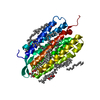 9f9bC 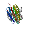 9f9cC  9f9dC  9f9eC  9f9fC  9f9gC  9f9iC  9f9jC C: citing same article ( |
|---|---|
| Similar structure data |
-Links
 PDBj
PDBj


- Assembly
| Deposited unit | 
| ||||||||
|---|---|---|---|---|---|---|---|---|---|
| 1 |
| ||||||||
| Unit cell |
|
-Components
| #1: Protein | Mass: 25261.807 Da / Num. of mol.: 1 Source method: isolated from a genetically manipulated source Source: (gene. exp.) Halobacterium salinarum (Halophile) / Gene: bop, VNG_1467G / Production host:  | ||||||||||
|---|---|---|---|---|---|---|---|---|---|---|---|
| #2: Chemical | ChemComp-LI1 / 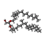  Mass: 639.130 Da / Num. of mol.: 12 / Source method: obtained synthetically / Formula: C42H86O3 Mass: 639.130 Da / Num. of mol.: 12 / Source method: obtained synthetically / Formula: C42H86O3#3: Chemical | ChemComp-OLC / (   Mass: 356.540 Da / Num. of mol.: 4 / Source method: obtained synthetically / Formula: C21H40O4 Mass: 356.540 Da / Num. of mol.: 4 / Source method: obtained synthetically / Formula: C21H40O4#4: Chemical | ChemComp-RET / |   Mass: 284.436 Da / Num. of mol.: 1 Mass: 284.436 Da / Num. of mol.: 1Source method: isolated from a genetically manipulated source Formula: C20H28O / Source: (gene. exp.) Halobacterium salinarum (Halophile) / Gene: bop, VNG_1467G / Production host: #5: Water | ChemComp-HOH / |  Mass: 18.015 Da / Num. of mol.: 40 / Source method: isolated from a natural source / Formula: H2O Mass: 18.015 Da / Num. of mol.: 40 / Source method: isolated from a natural source / Formula: H2OHas ligand of interest | Y | Has protein modification | Y | |
-Experimental details
-Experiment
| Experiment | Method: X-RAY DIFFRACTION / Number of used crystals: 1 |
|---|
- Sample preparation
| Crystal | Density Matthews: 2.5 Å3/Da / Density % sol: 50.85 % |
|---|---|
| Crystal grow | Temperature: 294 K / Method: lipidic cubic phase / pH: 5.6 / Details: 100 mM Na/K Phosphate buffer pH 5.6 30 % PEG 2000 |
-Data collection
| Diffraction | Mean temperature: 298 K / Serial crystal experiment: N |
|---|---|
| Diffraction source | Source: FREE ELECTRON LASER / Site: SLAC LCLS  / Beamline: CXI / Wavelength: 1.3 Å / Beamline: CXI / Wavelength: 1.3 Å |
| Detector | Type: CS-PAD CXI-1 / Detector: PIXEL / Date: Jun 30, 2017 |
| Radiation | Protocol: SINGLE WAVELENGTH / Monochromatic (M) / Laue (L): M / Scattering type: x-ray |
| Radiation wavelength | Wavelength: 1.3 Å / Relative weight: 1 |
| Reflection | Resolution: 1.55→19.35 Å / Num. obs: 35453 / % possible obs: 100 % / Redundancy: 194 % / CC star: 0.992 / R split: 0.083 / Net I/σ(I): 9.19 |
| Reflection shell | Resolution: 1.55→1.6 Å / Mean I/σ(I) obs: 1.19 / Num. unique obs: 3534 / CC star: 0.729 / R split: 0.9952 / % possible all: 100 |
- Processing
| Software |
| ||||||||||||||||||||||||||||||||||||||||||||||||||||||||||||||||||||||||||||||||||||||||||||||||||||||||||||||||||||||||||||||||||||||||||||||||||||||||||||||||||||||||||||||||||||||||||||||||||||||||||||||||||||||||||||||||||||||||||||||||||||||||||
|---|---|---|---|---|---|---|---|---|---|---|---|---|---|---|---|---|---|---|---|---|---|---|---|---|---|---|---|---|---|---|---|---|---|---|---|---|---|---|---|---|---|---|---|---|---|---|---|---|---|---|---|---|---|---|---|---|---|---|---|---|---|---|---|---|---|---|---|---|---|---|---|---|---|---|---|---|---|---|---|---|---|---|---|---|---|---|---|---|---|---|---|---|---|---|---|---|---|---|---|---|---|---|---|---|---|---|---|---|---|---|---|---|---|---|---|---|---|---|---|---|---|---|---|---|---|---|---|---|---|---|---|---|---|---|---|---|---|---|---|---|---|---|---|---|---|---|---|---|---|---|---|---|---|---|---|---|---|---|---|---|---|---|---|---|---|---|---|---|---|---|---|---|---|---|---|---|---|---|---|---|---|---|---|---|---|---|---|---|---|---|---|---|---|---|---|---|---|---|---|---|---|---|---|---|---|---|---|---|---|---|---|---|---|---|---|---|---|---|---|---|---|---|---|---|---|---|---|---|---|---|---|---|---|---|---|---|---|---|---|---|---|---|---|---|---|---|---|---|---|---|---|
| Refinement | Method to determine structure: MOLECULAR REPLACEMENT / Resolution: 1.66→16.02 Å / SU ML: 0.34 / Cross valid method: FREE R-VALUE / σ(F): 0 / Phase error: 34.09 / Stereochemistry target values: ML
| ||||||||||||||||||||||||||||||||||||||||||||||||||||||||||||||||||||||||||||||||||||||||||||||||||||||||||||||||||||||||||||||||||||||||||||||||||||||||||||||||||||||||||||||||||||||||||||||||||||||||||||||||||||||||||||||||||||||||||||||||||||||||||
| Solvent computation | Shrinkage radii: 0.9 Å / VDW probe radii: 1.1 Å / Solvent model: FLAT BULK SOLVENT MODEL | ||||||||||||||||||||||||||||||||||||||||||||||||||||||||||||||||||||||||||||||||||||||||||||||||||||||||||||||||||||||||||||||||||||||||||||||||||||||||||||||||||||||||||||||||||||||||||||||||||||||||||||||||||||||||||||||||||||||||||||||||||||||||||
| Refinement step | Cycle: LAST / Resolution: 1.66→16.02 Å
| ||||||||||||||||||||||||||||||||||||||||||||||||||||||||||||||||||||||||||||||||||||||||||||||||||||||||||||||||||||||||||||||||||||||||||||||||||||||||||||||||||||||||||||||||||||||||||||||||||||||||||||||||||||||||||||||||||||||||||||||||||||||||||
| Refine LS restraints |
| ||||||||||||||||||||||||||||||||||||||||||||||||||||||||||||||||||||||||||||||||||||||||||||||||||||||||||||||||||||||||||||||||||||||||||||||||||||||||||||||||||||||||||||||||||||||||||||||||||||||||||||||||||||||||||||||||||||||||||||||||||||||||||
| LS refinement shell |
| ||||||||||||||||||||||||||||||||||||||||||||||||||||||||||||||||||||||||||||||||||||||||||||||||||||||||||||||||||||||||||||||||||||||||||||||||||||||||||||||||||||||||||||||||||||||||||||||||||||||||||||||||||||||||||||||||||||||||||||||||||||||||||
| Refinement TLS params. | Method: refined / Refine-ID: X-RAY DIFFRACTION
| ||||||||||||||||||||||||||||||||||||||||||||||||||||||||||||||||||||||||||||||||||||||||||||||||||||||||||||||||||||||||||||||||||||||||||||||||||||||||||||||||||||||||||||||||||||||||||||||||||||||||||||||||||||||||||||||||||||||||||||||||||||||||||
| Refinement TLS group |
|
