 Movie
Movie Controller

[English] 日本語
 Yorodumi
- PDB-9f4b: Pre-assembled baseplate cup of Klebsiella phage KP1 variant vB_Kp... -
+ Open data

- Basic information
| Entry | Database: PDB / ID: 9f4b | |||||||||||||||||||||||||||||||||
|---|---|---|---|---|---|---|---|---|---|---|---|---|---|---|---|---|---|---|---|---|---|---|---|---|---|---|---|---|---|---|---|---|---|---|
| Title | Pre-assembled baseplate cup of Klebsiella phage KP1 variant vB_Kpn_Lilla1 | |||||||||||||||||||||||||||||||||
 Components Components |
| |||||||||||||||||||||||||||||||||
 Keywords Keywords | VIRUS / Baseplate cup / baseplate wedge / fibers | |||||||||||||||||||||||||||||||||
| Function / homology |  Function and homology information Function and homology informationsymbiont entry into host cell via disruption of host cell wall peptidoglycan / virus tail, baseplate / viral tail assembly / symbiont entry into host cell via disruption of host cell envelope / viral release from host cell / peptidoglycan catabolic process / cell wall macromolecule catabolic process / lysozyme / lysozyme activity / killing of cells of another organism ...symbiont entry into host cell via disruption of host cell wall peptidoglycan / virus tail, baseplate / viral tail assembly / symbiont entry into host cell via disruption of host cell envelope / viral release from host cell / peptidoglycan catabolic process / cell wall macromolecule catabolic process / lysozyme / lysozyme activity / killing of cells of another organism / defense response to bacterium / structural molecule activity Similarity search - Function | |||||||||||||||||||||||||||||||||
| Biological species |  Klebsiella phage KP1 (virus) Klebsiella phage KP1 (virus) | |||||||||||||||||||||||||||||||||
| Method | ELECTRON MICROSCOPY / single particle reconstruction / cryo EM / Resolution: 3.36 Å | |||||||||||||||||||||||||||||||||
 Authors Authors | Orlova, E.V. / Isupov, M.N. | |||||||||||||||||||||||||||||||||
| Funding support | 1items
| |||||||||||||||||||||||||||||||||
 Citation Citation | Journal: To Be Published Title: Baseplate cup of Klebsiella phage KP1 variant vB_Kpn_Lilla1 Authors: Orlova, E.V. / Isupov, M.N. | |||||||||||||||||||||||||||||||||
| History |
|
- Structure visualization
| Structure viewer | Molecule: MolmilJmol/JSmol |
|---|
- Downloads & links
-Download
| PDBx/mmCIF format | 9f4b.cif.gz | 10 MB | Display | PDBx/mmCIF format |
|---|---|---|---|---|
| PDB format | pdb9f4b.ent.gz | Display | PDB format | |
| PDBx/mmJSON format | 9f4b.json.gz | Tree view | PDBx/mmJSON format | |
| Others |  Other downloads Other downloads |
-Validation report
| Arichive directory | https://data.pdbj.org/pub/pdb/validation_reports/f4/9f4bftp://data.pdbj.org/pub/pdb/validation_reports/f4/9f4b | HTTPS FTP |
|---|
-Related structure data
| Related structure data |  50187MC 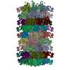 9ev2C  9f4aC M: map data used to model this data C: citing same article ( |
|---|---|
| Similar structure data |

-Links
 PDBj
PDBj


- Assembly
| Deposited unit | 
|
|---|---|
| 1 |
|
-Components
-Baseplate wedge ... , 7 types, 90 molecules ATAVAXASAUAWANAPARAMAOAQAHAJALAGAIAKAYA2AZA0A1A3AdAcAbAaAeAf...
| #1: Protein | Mass: 73689.242 Da / Num. of mol.: 12 / Source method: isolated from a natural source / Source: (natural) Klebsiella phage KP1 (virus) / References: UniProt: A0A2K9V5R4#4: Protein | Mass: 24693.420 Da / Num. of mol.: 6 / Source method: isolated from a natural source / Source: (natural) Klebsiella phage KP1 (virus) / References: UniProt: A0A2K9V5R5#7: Protein | Mass: 119645.719 Da / Num. of mol.: 6 / Source method: isolated from a natural source / Source: (natural) Klebsiella phage KP1 (virus) / References: UniProt: A0A2K9V5T9#8: Protein | Mass: 38704.387 Da / Num. of mol.: 12 / Source method: isolated from a natural source / Source: (natural) Klebsiella phage KP1 (virus) / References: UniProt: A0A2Z4QAY9#9: Protein | Mass: 32380.195 Da / Num. of mol.: 18 / Source method: isolated from a natural source / Source: (natural) Klebsiella phage KP1 (virus) / References: UniProt: A0A2K9V5S1#10: Protein | Mass: 66712.070 Da / Num. of mol.: 18 / Source method: isolated from a natural source / Source: (natural) Klebsiella phage KP1 (virus) / References: UniProt: A0A2K9V5S8#11: Protein | Mass: 24534.469 Da / Num. of mol.: 18 / Source method: isolated from a natural source / Source: (natural) Klebsiella phage KP1 (virus) / References: UniProt: A0A2K9V5T5 |
|---|
-Protein , 9 types, 58 molecules BLBNBPBKBMBOBQBRBSBTBUBVBBBCBDBAFGFIFHFPFRFQFYFaFZFhFjFiFqFs...
| #2: Protein | Mass: 38263.664 Da / Num. of mol.: 6 / Source method: isolated from a natural source / Source: (natural) Klebsiella phage KP1 (virus) / References: UniProt: A0A2K9V618#3: Protein | Mass: 33716.129 Da / Num. of mol.: 6 / Source method: isolated from a natural source / Source: (natural) Klebsiella phage KP1 (virus) / References: UniProt: A0A7I8V4E3#5: Protein | Mass: 63317.457 Da / Num. of mol.: 3 / Source method: isolated from a natural source / Source: (natural) Klebsiella phage KP1 (virus) / References: UniProt: A0A976LXP5, lysozyme#6: Protein | | Mass: 10096.360 Da / Num. of mol.: 1 / Source method: isolated from a natural source / Source: (natural) Klebsiella phage KP1 (virus) / References: UniProt: A0A2K9V5W6#12: Protein | Mass: 48092.477 Da / Num. of mol.: 18 / Source method: isolated from a natural source / Source: (natural) Klebsiella phage KP1 (virus) / References: UniProt: A0A7R8MRX7#13: Protein | Mass: 18633.738 Da / Num. of mol.: 12 / Source method: isolated from a natural source / Source: (natural) Klebsiella phage KP1 (virus) / References: UniProt: A0A2K9V5T6#14: Protein | Mass: 15382.360 Da / Num. of mol.: 6 / Source method: isolated from a natural source / Source: (natural) Klebsiella phage KP1 (virus) / References: UniProt: A0A0K1Y5J2#15: Protein | Mass: 43526.016 Da / Num. of mol.: 3 / Source method: isolated from a natural source / Source: (natural) Klebsiella phage KP1 (virus) / References: UniProt: A0A7T3TJP2#16: Protein | Mass: 64127.020 Da / Num. of mol.: 3 / Source method: isolated from a natural source / Source: (natural) Klebsiella phage KP1 (virus) / References: UniProt: A0A2K9V5Z4 |
|---|
-Non-polymers , 3 types, 13 molecules 


| #17: Chemical | ChemComp-CL /  Mass: 35.453 Da / Num. of mol.: 1 / Source method: obtained synthetically / Formula: Cl Mass: 35.453 Da / Num. of mol.: 1 / Source method: obtained synthetically / Formula: Cl | ||
|---|---|---|---|
| #18: Chemical | ChemComp-K /  Mass: 39.098 Da / Num. of mol.: 6 / Source method: obtained synthetically / Formula: K / Feature type: SUBJECT OF INVESTIGATION Mass: 39.098 Da / Num. of mol.: 6 / Source method: obtained synthetically / Formula: K / Feature type: SUBJECT OF INVESTIGATION#19: Chemical | ChemComp-ZN /  Mass: 65.409 Da / Num. of mol.: 6 / Source method: obtained synthetically / Formula: Zn / Feature type: SUBJECT OF INVESTIGATION Mass: 65.409 Da / Num. of mol.: 6 / Source method: obtained synthetically / Formula: Zn / Feature type: SUBJECT OF INVESTIGATION |
-Details
| Has ligand of interest | Y |
|---|---|
| Has protein modification | Y |
-Experimental details
-Experiment
| Experiment | Method: ELECTRON MICROSCOPY |
|---|---|
| EM experiment | Aggregation state: PARTICLE / 3D reconstruction method: single particle reconstruction |
- Sample preparation
| Component | Name: vB_Kpn_Lilla1 / Type: VIRUS / Entity ID: #1-#16 / Source: NATURAL |
|---|---|
| Molecular weight | Experimental value: NO |
| Source (natural) | Organism: vB_Kpn_Lilla1 (virus) |
| Details of virus | Empty: NO / Enveloped: NO / Isolate: OTHER / Type: VIRION |
| Natural host | Organism: Klebsiella |
| Buffer solution | pH: 7.5 / Details: 100mM NaCl, 8mM MgSO4, 50mM Tris-HCl |
| Specimen | Embedding applied: NO / Shadowing applied: NO / Staining applied: NO / Vitrification applied: YES / Details: Klebsiella phage KP1 variant vB_Kpn_Lilla1 |
| Specimen support | Grid material: COPPER / Grid mesh size: 400 divisions/in. / Grid type: EMS Lacey Carbon |
| Vitrification | Instrument: FEI VITROBOT MARK IV / Cryogen name: ETHANE / Humidity: 92 % / Chamber temperature: 281 K |
- Electron microscopy imaging
| Experimental equipment |  Model: Titan Krios / Image courtesy: FEI Company |
|---|---|
| Microscopy | Model: TFS KRIOS Details: Alignment procedure: EPU Autocoma and Autosrigmate Residual tilt (if alignment procedure is coma free, mrad): 68 Software used to collect images EPU 2.4, Tomo 5.13 Film/CCD/Direct electron ...Details: Alignment procedure: EPU Autocoma and Autosrigmate Residual tilt (if alignment procedure is coma free, mrad): 68 Software used to collect images EPU 2.4, Tomo 5.13 Film/CCD/Direct electron detector model*: post GIF K3 |
| Electron gun | Electron source:  FIELD EMISSION GUN / Accelerating voltage: 300 kV / Illumination mode: FLOOD BEAM FIELD EMISSION GUN / Accelerating voltage: 300 kV / Illumination mode: FLOOD BEAM |
| Electron lens | Mode: BRIGHT FIELD / Nominal magnification: 81000 X / Calibrated magnification: 83505 X / Nominal defocus max: 2500 nm / Nominal defocus min: 500 nm / Cs: 2.7 mm / C2 aperture diameter: 100 µm / Alignment procedure: OTHER |
| Specimen holder | Cryogen: NITROGEN / Specimen holder model: FEI TITAN KRIOS AUTOGRID HOLDER / Temperature (max): 93.6 K / Temperature (min): 78.7 K / Residual tilt: 68 mradians |
| Image recording | Average exposure time: 2 sec. / Electron dose: 40 e/Å2 / Detector mode: COUNTING / Film or detector model: GATAN K3 (6k x 4k) / Num. of grids imaged: 4 |
| Image scans | Sampling size: 5 µm / Width: 4092 / Height: 5760 |
- Processing
| EM software |
| ||||||||||||||||||||||||||||||||||||||||||||||||
|---|---|---|---|---|---|---|---|---|---|---|---|---|---|---|---|---|---|---|---|---|---|---|---|---|---|---|---|---|---|---|---|---|---|---|---|---|---|---|---|---|---|---|---|---|---|---|---|---|---|
| CTF correction | Type: PHASE FLIPPING AND AMPLITUDE CORRECTION | ||||||||||||||||||||||||||||||||||||||||||||||||
| Particle selection | Num. of particles selected: 34529 | ||||||||||||||||||||||||||||||||||||||||||||||||
| Symmetry | Point symmetry: C3 (3 fold cyclic) | ||||||||||||||||||||||||||||||||||||||||||||||||
| 3D reconstruction | Resolution: 3.36 Å / Resolution method: FSC 0.143 CUT-OFF / Num. of particles: 34528 / Algorithm: FOURIER SPACE / Num. of class averages: 98 / Symmetry type: POINT | ||||||||||||||||||||||||||||||||||||||||||||||||
| Atomic model building | Protocol: FLEXIBLE FIT / Space: REAL |
