 Movie
Movie Controller

[English] 日本語
 Yorodumi
- PDB-9ev2: Tail tube and extended tail sheath tube of Klebsiella phage KP1 v... -
+ Open data

- Basic information
| Entry | Database: PDB / ID: 9ev2 | ||||||||||||||||||||||||
|---|---|---|---|---|---|---|---|---|---|---|---|---|---|---|---|---|---|---|---|---|---|---|---|---|---|
| Title | Tail tube and extended tail sheath tube of Klebsiella phage KP1 variant vB_Kpn_Lilla1 | ||||||||||||||||||||||||
 Components Components |
| ||||||||||||||||||||||||
 Keywords Keywords | VIRUS / Viral tail / sheath | ||||||||||||||||||||||||
| Function / homology |  Function and homology information Function and homology informationvirus tail, sheath / symbiont genome ejection through host cell envelope, contractile tail mechanism / structural molecule activity Similarity search - Function | ||||||||||||||||||||||||
| Biological species |  Klebsiella phage KP1 (virus) Klebsiella phage KP1 (virus) | ||||||||||||||||||||||||
| Method | ELECTRON MICROSCOPY / helical reconstruction / cryo EM / Resolution: 3.8 Å | ||||||||||||||||||||||||
 Authors Authors | Orlova, E.V. / Isupov, M.N. | ||||||||||||||||||||||||
| Funding support | 1items
| ||||||||||||||||||||||||
 Citation Citation | Journal: To Be Published Title: Baseplate cup of Klebsiella phage KP1 variant vB_Kpn_Lilla1 Authors: Orlova, E.V. / Isupov, M.N. | ||||||||||||||||||||||||
| History |
|
- Structure visualization
| Structure viewer | Molecule: MolmilJmol/JSmol |
|---|
- Downloads & links
-Download
| PDBx/mmCIF format | 9ev2.cif.gz | 7.8 MB | Display | PDBx/mmCIF format |
|---|---|---|---|---|
| PDB format | pdb9ev2.ent.gz | Display | PDB format | |
| PDBx/mmJSON format | 9ev2.json.gz | Tree view | PDBx/mmJSON format | |
| Others |  Other downloads Other downloads |
-Validation report
| Arichive directory | https://data.pdbj.org/pub/pdb/validation_reports/ev/9ev2ftp://data.pdbj.org/pub/pdb/validation_reports/ev/9ev2 | HTTPS FTP |
|---|
-Related structure data
| Related structure data | 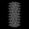 19992MC  9f4aC  9f4bC M: map data used to model this data C: citing same article ( |
|---|---|
| Similar structure data |


-Links
 PDBj
PDBj- Assembly
| Deposited unit | 
|
|---|---|
| 1 |
|
-Components
| #1: Protein | Mass: 71695.094 Da / Num. of mol.: 54 / Source method: isolated from a natural source / Source: (natural) Klebsiella phage KP1 (virus) / References: UniProt: A0A2K9V5S7#2: Protein | Mass: 18633.738 Da / Num. of mol.: 54 / Source method: isolated from a natural source / Source: (natural) Klebsiella phage KP1 (virus) / References: UniProt: A0A2K9V5T6Has protein modification | N | |
|---|
-Experimental details
-Experiment
| Experiment | Method: ELECTRON MICROSCOPY |
|---|---|
| EM experiment | Aggregation state: PARTICLE / 3D reconstruction method: helical reconstruction |
- Sample preparation
| Component | Name: Klebsiella phage KP1 / Type: VIRUS / Entity ID: all / Source: NATURAL |
|---|---|
| Source (natural) | Organism: Klebsiella phage KP1 (virus) |
| Details of virus | Empty: NO / Enveloped: NO / Isolate: OTHER / Type: VIRION |
| Buffer solution | pH: 7.3 |
| Specimen | Embedding applied: NO / Shadowing applied: NO / Staining applied: NO / Vitrification applied: YES / Details: Klebsiella phage KP1 variant vB_Kpn_Lilla1 |
| Specimen support | Grid material: COPPER / Grid mesh size: 400 divisions/in. / Grid type: EMS Lacey Carbon |
| Vitrification | Instrument: FEI VITROBOT MARK IV / Cryogen name: ETHANE / Humidity: 92 % / Chamber temperature: 281 K |
- Electron microscopy imaging
| Experimental equipment |  Model: Titan Krios / Image courtesy: FEI Company |
|---|---|
| Microscopy | Model: TFS KRIOS Details: Alignment procedure: EPU Autocoma and Autosrigmate Residual tilt (if alignment procedure is coma free, mrad): 68 Software used to collect images EPU 2.4, Tomo 5.13 Film/CCD/Direct electron ...Details: Alignment procedure: EPU Autocoma and Autosrigmate Residual tilt (if alignment procedure is coma free, mrad): 68 Software used to collect images EPU 2.4, Tomo 5.13 Film/CCD/Direct electron detector model*: post GIF K3 |
| Electron gun | Electron source:  FIELD EMISSION GUN / Accelerating voltage: 300 kV / Illumination mode: FLOOD BEAM FIELD EMISSION GUN / Accelerating voltage: 300 kV / Illumination mode: FLOOD BEAM |
| Electron lens | Mode: BRIGHT FIELD / Nominal magnification: 81000 X / Calibrated magnification: 83505 X / Nominal defocus max: 2500 nm / Nominal defocus min: 500 nm / Cs: 2.7 mm / C2 aperture diameter: 100 µm / Alignment procedure: OTHER |
| Specimen holder | Cryogen: NITROGEN / Specimen holder model: FEI TITAN KRIOS AUTOGRID HOLDER / Temperature (max): 93.6 K / Temperature (min): 78.7 K / Residual tilt: 68 mradians |
| Image recording | Electron dose: 40 e/Å2 / Detector mode: COUNTING / Film or detector model: GATAN K3 (6k x 4k) |
- Processing
| EM software |
| ||||||||||||||||||||||||||||||||||||||||||||||||||||||||
|---|---|---|---|---|---|---|---|---|---|---|---|---|---|---|---|---|---|---|---|---|---|---|---|---|---|---|---|---|---|---|---|---|---|---|---|---|---|---|---|---|---|---|---|---|---|---|---|---|---|---|---|---|---|---|---|---|---|
| CTF correction | Type: PHASE FLIPPING AND AMPLITUDE CORRECTION | ||||||||||||||||||||||||||||||||||||||||||||||||||||||||
| Helical symmerty | Angular rotation/subunit: 16.93 ° / Axial rise/subunit: 40.22 Å / Axial symmetry: C6 | ||||||||||||||||||||||||||||||||||||||||||||||||||||||||
| Particle selection | Num. of particles selected: 40161 | ||||||||||||||||||||||||||||||||||||||||||||||||||||||||
| 3D reconstruction | Resolution: 3.8 Å / Resolution method: FSC 0.143 CUT-OFF / Num. of particles: 7017 / Algorithm: FOURIER SPACE / Symmetry type: HELICAL | ||||||||||||||||||||||||||||||||||||||||||||||||||||||||
| Atomic model building | Protocol: FLEXIBLE FIT |
