+ データを開く
- 基本情報
基本情報
| 登録情報 | データベース: PDB / ID: 9f48 | |||||||||
|---|---|---|---|---|---|---|---|---|---|---|
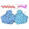
| タイトル | KS + AT di-domain of polyketide synthase 13 in Mycobacterium tuberculosis | |||||||||
 要素 要素 | Polyketide synthase Pks13 | |||||||||
 キーワード キーワード | BIOSYNTHETIC PROTEIN / Mycolic acid synthesis / Claisen condensation / cell wall synthesis | |||||||||
| 機能・相同性 |  機能・相同性情報 機能・相同性情報DIM/DIP cell wall layer assembly / fatty acid synthase activity / phosphopantetheine binding / 3-oxoacyl-[acyl-carrier-protein] synthase activity / 転移酵素; アシル基を移すもの; アミノアシル基以外のアシル基を移すもの / fatty acid biosynthetic process / cytoplasm 類似検索 - 分子機能 | |||||||||
| 生物種 |  Mycobacterium tuberculosis H37Rv (結核菌) Mycobacterium tuberculosis H37Rv (結核菌) | |||||||||
| 手法 | 電子顕微鏡法 / 単粒子再構成法 / クライオ電子顕微鏡法 / 解像度: 3.4 Å | |||||||||
 データ登録者 データ登録者 | Johnston, H.E. / Futterer, K. | |||||||||
| 資金援助 |  英国, 2件 英国, 2件
| |||||||||
 引用 引用 |  ジャーナル: Microbiology (Reading) / 年: 2024 ジャーナル: Microbiology (Reading) / 年: 2024タイトル: Cryo-electron microscopy structure of the di-domain core of polyketide synthase 13, essential for mycobacterial mycolic acid synthesis. 著者: Hannah E Johnston / Sarah M Batt / Alistair K Brown / Christos G Savva / Gurdyal S Besra / Klaus Fütterer /  要旨: Mycobacteria are known for their complex cell wall, which comprises layers of peptidoglycan, polysaccharides and unusual fatty acids known as mycolic acids that form their unique outer membrane. ...Mycobacteria are known for their complex cell wall, which comprises layers of peptidoglycan, polysaccharides and unusual fatty acids known as mycolic acids that form their unique outer membrane. Polyketide synthase 13 (Pks13) of , the bacterial organism causing tuberculosis, catalyses the last step of mycolic acid synthesis prior to export to and assembly in the cell wall. Due to its essentiality, Pks13 is a target for several novel anti-tubercular inhibitors, but its 3D structure and catalytic reaction mechanism remain to be fully elucidated. Here, we report the molecular structure of the catalytic core domains of Pks13 (Mt-Pks13), determined by transmission cryo-electron microscopy (cryoEM) to a resolution of 3.4 Å. We observed a homodimeric assembly comprising the ketoacyl synthase (KS) domain at the centre, mediating dimerization, and the acyltransferase (AT) domains protruding in opposite directions from the central KS domain dimer. In addition to the KS-AT di-domains, the cryoEM map includes features not covered by the di-domain structural model that we predicted to contain a dimeric domain similar to dehydratases, yet likely lacking catalytic function. Analytical ultracentrifugation data indicate a pH-dependent equilibrium between monomeric and dimeric assembly states, while comparison with the previously determined structures of Pks13 indicates architectural flexibility. Combining the experimentally determined structure with modelling in AlphaFold2 suggests a structural scaffold with a relatively stable dimeric core, which combines with considerable conformational flexibility to facilitate the successive steps of the Claisen-type condensation reaction catalysed by Pks13. | |||||||||
| 履歴 |
|
- 構造の表示
構造の表示
| 構造ビューア | 分子:  Molmil Molmil Jmol/JSmol Jmol/JSmol |
|---|
- ダウンロードとリンク
- ダウンロード
| PDBx/mmCIF形式 |  9f48.cif.gz 9f48.cif.gz | 459 KB | 表示 |  PDBx/mmCIF形式 PDBx/mmCIF形式 |
|---|---|---|---|---|
| PDB形式 |  pdb9f48.ent.gz pdb9f48.ent.gz | 287.8 KB | 表示 |  PDB形式 PDB形式 |
| PDBx/mmJSON形式 |  9f48.json.gz 9f48.json.gz | ツリー表示 |  PDBx/mmJSON形式 PDBx/mmJSON形式 | |
| その他 |  その他のダウンロード その他のダウンロード |
-検証レポート
| アーカイブディレクトリ |  https://data.pdbj.org/pub/pdb/validation_reports/f4/9f48 https://data.pdbj.org/pub/pdb/validation_reports/f4/9f48 ftp://data.pdbj.org/pub/pdb/validation_reports/f4/9f48 ftp://data.pdbj.org/pub/pdb/validation_reports/f4/9f48 | HTTPS FTP |
|---|
-関連構造データ
| 関連構造データ |  50185MC M: このデータのモデリングに利用したマップデータ C: 同じ文献を引用 ( |
|---|---|
| 類似構造データ | 類似検索 - 機能・相同性  F&H 検索 F&H 検索 |
- リンク
リンク
- 集合体
集合体
| 登録構造単位 | 
|
|---|---|
| 1 |
|
- 要素
要素
| #1: タンパク質 | 分子量: 186642.188 Da / 分子数: 2 / 由来タイプ: 組換発現 由来: (組換発現)  Mycobacterium tuberculosis H37Rv (結核菌) Mycobacterium tuberculosis H37Rv (結核菌)遺伝子: pks13, Rv3800c / 発現宿主:  参照: UniProt: I6X8D2, 転移酵素; アシル基を移すもの; アミノアシル基以外のアシル基を移すもの Has protein modification | N | |
|---|
-実験情報
-実験
| 実験 | 手法: 電子顕微鏡法 |
|---|---|
| EM実験 | 試料の集合状態: PARTICLE / 3次元再構成法: 単粒子再構成法 |
- 試料調製
試料調製
| 構成要素 | 名称: Subunit of Pks13 / タイプ: COMPLEX / Entity ID: all / 由来: RECOMBINANT |
|---|---|
| 分子量 | 値: 0.187 MDa / 実験値: YES |
| 由来(天然) | 生物種:  |
| 由来(組換発現) | 生物種:  |
| 緩衝液 | pH: 7.9 詳細: 20 mM Tris-HCl pH 7.9, 50 mM NaCl, 2.5 mM beta-mercaptoethanol |
| 緩衝液成分 | 濃度: 20 mM / 名称: Tris / 式: C4H11NO3 |
| 試料 | 濃度: 0.9 mg/ml / 包埋: NO / シャドウイング: NO / 染色: NO / 凍結: YES |
| 試料支持 | グリッドの材料: GOLD / グリッドのタイプ: Quantifoil R1.2/1.3 |
| 急速凍結 | 装置: FEI VITROBOT MARK IV / 凍結剤: ETHANE / 湿度: 100 % / 凍結前の試料温度: 291 K |
- 電子顕微鏡撮影
| 実験機器 |  モデル: Titan Krios / 画像提供: FEI Company |
|---|---|
| 顕微鏡 | モデル: FEI TITAN KRIOS |
| 電子銃 | 電子線源:  FIELD EMISSION GUN / 加速電圧: 300 kV / 照射モード: FLOOD BEAM FIELD EMISSION GUN / 加速電圧: 300 kV / 照射モード: FLOOD BEAM |
| 電子レンズ | モード: OTHER / 最大 デフォーカス(公称値): 2700 nm / 最小 デフォーカス(公称値): 1500 nm / Cs: 2.7 mm / C2レンズ絞り径: 100 µm |
| 試料ホルダ | 試料ホルダーモデル: FEI TITAN KRIOS AUTOGRID HOLDER |
| 撮影 | 平均露光時間: 3 sec. / 電子線照射量: 16.5 e/Å2 / フィルム・検出器のモデル: GATAN K3 (6k x 4k) / 撮影したグリッド数: 1 / 実像数: 1412 / 詳細: movie mode |
- 解析
解析
| EMソフトウェア | 名称: PHENIX / バージョン: 1.21.1_5286 / カテゴリ: モデル精密化 | ||||||||||||||||||||||||
|---|---|---|---|---|---|---|---|---|---|---|---|---|---|---|---|---|---|---|---|---|---|---|---|---|---|
| CTF補正 | タイプ: NONE | ||||||||||||||||||||||||
| 粒子像の選択 | 選択した粒子像数: 310055 | ||||||||||||||||||||||||
| 3次元再構成 | 解像度: 3.4 Å / 解像度の算出法: OTHER / 粒子像の数: 168566 / 対称性のタイプ: POINT | ||||||||||||||||||||||||
| 精密化 | 交差検証法: NONE 立体化学のターゲット値: GeoStd + Monomer Library + CDL v1.2 | ||||||||||||||||||||||||
| 原子変位パラメータ | Biso mean: 70.54 Å2 | ||||||||||||||||||||||||
| 拘束条件 |
|
 ムービー
ムービー コントローラー


 PDBj
PDBj


