 Movie
Movie Controller

[English] 日本語
 Yorodumi
- PDB-9f1u: Cryo-EM structure of the I923V MDA5-dsRNA filament with ADP-AlF4 ... -
+ Open data

- Basic information
| Entry | Database: PDB / ID: 9f1u | |||||||||||||||||||||||||||
|---|---|---|---|---|---|---|---|---|---|---|---|---|---|---|---|---|---|---|---|---|---|---|---|---|---|---|---|---|
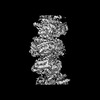
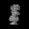
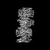
| Title | Cryo-EM structure of the I923V MDA5-dsRNA filament with ADP-AlF4 bound and 81-degree helical twist | |||||||||||||||||||||||||||
 Components Components |
| |||||||||||||||||||||||||||
 Keywords Keywords | IMMUNE SYSTEM / PROTEIN-RNA COMPLEX / HELICAL FILAMENT / ATPASE / INNATE IMMUNE RECEPTOR | |||||||||||||||||||||||||||
| Function / homology |  Function and homology information Function and homology informationMDA-5 signaling pathway / positive regulation of response to cytokine stimulus / Ub-specific processing proteases / mRNA transcription / negative regulation of viral genome replication / type I interferon-mediated signaling pathway / cellular response to exogenous dsRNA / pattern recognition receptor activity / protein complex oligomerization / positive regulation of interferon-alpha production ...MDA-5 signaling pathway / positive regulation of response to cytokine stimulus / Ub-specific processing proteases / mRNA transcription / negative regulation of viral genome replication / type I interferon-mediated signaling pathway / cellular response to exogenous dsRNA / pattern recognition receptor activity / protein complex oligomerization / positive regulation of interferon-alpha production / protein sumoylation / ribonucleoprotein complex binding / positive regulation of interferon-beta production / antiviral innate immune response / cellular response to virus / positive regulation of interleukin-6 production / response to virus / positive regulation of tumor necrosis factor production / double-stranded RNA binding / defense response to virus / RNA helicase activity / single-stranded RNA binding / RNA helicase / protein domain specific binding / innate immune response / ATP hydrolysis activity / mitochondrion / DNA binding / RNA binding / zinc ion binding / ATP binding / identical protein binding / nucleus / cytoplasm Similarity search - Function | |||||||||||||||||||||||||||
| Biological species |  | |||||||||||||||||||||||||||
| Method | ELECTRON MICROSCOPY / helical reconstruction / cryo EM / Resolution: 3.67 Å | |||||||||||||||||||||||||||
 Authors Authors | Singh, R. / Herrero del Valle, A. / Modis, Y. | |||||||||||||||||||||||||||
| Funding support |  United Kingdom, United Kingdom,  France, 4items France, 4items
| |||||||||||||||||||||||||||
 Citation Citation | Journal: Cell Rep / Year: 2025 Title: Molecular basis of autoimmune disease protection by MDA5 variants. Authors: Rahul Singh / Joe D Joiner / Alba Herrero Del Valle / Marleen Zwaagstra / Ida Jobe / Brian J Ferguson / Frank J M van Kuppeveld / Yorgo Modis /  Abstract: MDA5 recognizes double-stranded RNA (dsRNA) from viruses and retroelements. Cooperative filament formation and ATP-dependent proofreading confer MDA5 with the necessary sensitivity and specificity ...MDA5 recognizes double-stranded RNA (dsRNA) from viruses and retroelements. Cooperative filament formation and ATP-dependent proofreading confer MDA5 with the necessary sensitivity and specificity for dsRNA. Many MDA5 genetic variants are associated with protection from autoimmune disease while increasing the risk of infection and chronic inflammation. How these variants affect RNA sensing remains unclear. Here, we determine the consequences of autoimmune-protective variants on the molecular structure and activities of MDA5. Rare variants E627 and I923V reduce the interferon response to picornavirus infection. E627 does not bind RNA. I923V is ATPase hyperactive, causing premature dissociation from dsRNA. Cryoelectron microscopy (cryo-EM) structures of MDA5 I923V bound to dsRNA at different stages of ATP hydrolysis reveal smaller RNA binding interfaces, leading to excessive proofreading activity. Variants R843H and T946A, which are genetically linked and cause mild phenotypes, did not affect cytokine induction, suggesting an indirect disease mechanism. In conclusion, autoimmune-protective MDA5 variants dampen MDA5-dependent signaling via multiple mechanisms. #1: Journal: Biorxiv / Year: 2024Title: Molecular basis of autoimmune disease protection by MDA5 variants Authors: Singh, R. / Herrero del Valle, A. / Joiner, J.D. / Zwaagstra, M. / Ferguson, B.J. / van Kuppeveld, F.J.M. / Modis, Y. | |||||||||||||||||||||||||||
| History |
|
- Structure visualization
| Structure viewer | Molecule: MolmilJmol/JSmol |
|---|
- Downloads & links
-Download
| PDBx/mmCIF format | 9f1u.cif.gz | 270.3 KB | Display | PDBx/mmCIF format |
|---|---|---|---|---|
| PDB format | pdb9f1u.ent.gz | 206.1 KB | Display | PDB format |
| PDBx/mmJSON format | 9f1u.json.gz | Tree view | PDBx/mmJSON format | |
| Others |  Other downloads Other downloads |
-Validation report
| Arichive directory | https://data.pdbj.org/pub/pdb/validation_reports/f1/9f1uftp://data.pdbj.org/pub/pdb/validation_reports/f1/9f1u | HTTPS FTP |
|---|
-Related structure data
| Related structure data | 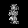 50136MC 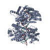 9f0jC 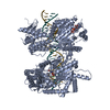 9f20C 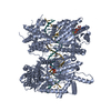 9f2lC 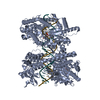 9f2wC 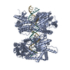 9f3pC C: citing same article ( M: map data used to model this data |
|---|---|
| Similar structure data |
-Links
 PDBj
PDBj


- Assembly
| Deposited unit | 
|
|---|---|
| 1 | 
|
-Components
-Protein , 1 types, 1 molecules A
| #1: Protein | Mass: 116683.062 Da / Num. of mol.: 1 / Mutation: I923V Source method: isolated from a genetically manipulated source Details: N-terminal hexahistidine tag and TEV cleavage site / Source: (gene. exp.)  |
|---|
-RNA chain , 2 types, 2 molecules XZ
| #2: RNA chain | Mass: 4548.812 Da / Num. of mol.: 1 / Source method: obtained synthetically / Details: In vitro transcribed RNA / Source: (synth.) |
|---|---|
| #3: RNA chain | Mass: 4376.604 Da / Num. of mol.: 1 / Source method: obtained synthetically / Details: In vitro transcribed RNA / Source: (synth.) |
-Non-polymers , 3 types, 3 molecules 


| #4: Chemical | ChemComp-ADP /  Mass: 427.201 Da / Num. of mol.: 1 / Source method: obtained synthetically / Formula: C10H15N5O10P2 / Feature type: SUBJECT OF INVESTIGATION / Comment: ADP, energy-carrying molecule*YM Mass: 427.201 Da / Num. of mol.: 1 / Source method: obtained synthetically / Formula: C10H15N5O10P2 / Feature type: SUBJECT OF INVESTIGATION / Comment: ADP, energy-carrying molecule*YM |
|---|---|
| #5: Chemical | ChemComp-ALF /  Mass: 102.975 Da / Num. of mol.: 1 / Source method: obtained synthetically / Formula: AlF4 / Feature type: SUBJECT OF INVESTIGATION Mass: 102.975 Da / Num. of mol.: 1 / Source method: obtained synthetically / Formula: AlF4 / Feature type: SUBJECT OF INVESTIGATION |
| #6: Chemical | ChemComp-ZN /  Mass: 65.409 Da / Num. of mol.: 1 / Source method: obtained synthetically / Formula: Zn Mass: 65.409 Da / Num. of mol.: 1 / Source method: obtained synthetically / Formula: Zn |
-Details
| Has ligand of interest | Y |
|---|---|
| Has protein modification | N |
-Experimental details
-Experiment
| Experiment | Method: ELECTRON MICROSCOPY |
|---|---|
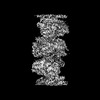
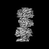
| EM experiment | Aggregation state: FILAMENT / 3D reconstruction method: helical reconstruction |
- Sample preparation
| Component |
| ||||||||||||||||||||||||||||||
|---|---|---|---|---|---|---|---|---|---|---|---|---|---|---|---|---|---|---|---|---|---|---|---|---|---|---|---|---|---|---|---|
| Molecular weight | Value: 28.03 kDa/nm / Experimental value: YES | ||||||||||||||||||||||||||||||
| Source (natural) |
| ||||||||||||||||||||||||||||||
| Source (recombinant) |
| ||||||||||||||||||||||||||||||
| Buffer solution | pH: 7.7 | ||||||||||||||||||||||||||||||
| Buffer component |
| ||||||||||||||||||||||||||||||
| Specimen | Conc.: 0.5 mg/ml / Embedding applied: NO / Shadowing applied: NO / Staining applied: NO / Vitrification applied: YES Details: 1 mg/ml MDA5 protein was incubated with 0.05 mg/ml 1-kb dsRNA on ice for 2 min. Samples were diluted twofold with buffer and applied to grids. | ||||||||||||||||||||||||||||||
| Specimen support | Details: Edwards 12E6/531 glow discharger at 30 mA / Grid material: GOLD / Grid mesh size: 300 divisions/in. / Grid type: Quantifoil R1.2/1.3 | ||||||||||||||||||||||||||||||
| Vitrification | Instrument: FEI VITROBOT MARK IV / Cryogen name: ETHANE / Humidity: 100 % / Chamber temperature: 277 K / Details: 2-4 s blotting, 15 s wait time |
- Electron microscopy imaging
| Experimental equipment |  Model: Titan Krios / Image courtesy: FEI Company |
|---|---|
| Microscopy | Model: FEI TITAN KRIOS |
| Electron gun | Electron source:  FIELD EMISSION GUN / Accelerating voltage: 300 kV / Illumination mode: FLOOD BEAM FIELD EMISSION GUN / Accelerating voltage: 300 kV / Illumination mode: FLOOD BEAM |
| Electron lens | Mode: DARK FIELD / Nominal magnification: 105000 X / Nominal defocus max: 2500 nm / Nominal defocus min: 500 nm / Cs: 2.7 mm / Alignment procedure: ZEMLIN TABLEAU |
| Specimen holder | Cryogen: NITROGEN / Specimen holder model: FEI TITAN KRIOS AUTOGRID HOLDER |
| Image recording | Electron dose: 44 e/Å2 / Film or detector model: GATAN K3 BIOQUANTUM (6k x 4k) |
| EM imaging optics | Energyfilter name: GIF Bioquantum / Energyfilter slit width: 20 eV |
- Processing
| EM software |
| ||||||||||||||||||||||||||||||||||||||||
|---|---|---|---|---|---|---|---|---|---|---|---|---|---|---|---|---|---|---|---|---|---|---|---|---|---|---|---|---|---|---|---|---|---|---|---|---|---|---|---|---|---|
| CTF correction | Type: PHASE FLIPPING AND AMPLITUDE CORRECTION | ||||||||||||||||||||||||||||||||||||||||
| Helical symmerty | Angular rotation/subunit: 80.71 ° / Axial rise/subunit: 43.86 Å / Axial symmetry: C1 | ||||||||||||||||||||||||||||||||||||||||
| Particle selection | Num. of particles selected: 1406812 | ||||||||||||||||||||||||||||||||||||||||
| 3D reconstruction | Resolution: 3.67 Å / Resolution method: FSC 0.143 CUT-OFF / Num. of particles: 347343 / Symmetry type: HELICAL | ||||||||||||||||||||||||||||||||||||||||
| Atomic model building | B value: 90 / Protocol: RIGID BODY FIT / Space: REAL / Target criteria: Cross-correlation coefficient Details: Initial local fitting with Fit-in-Map in Chimera followed by real space refinement in Phenix. | ||||||||||||||||||||||||||||||||||||||||
| Atomic model building | PDB-ID: 7NIQ Pdb chain-ID: A / Accession code: 7NIQ / Source name: PDB / Type: experimental model | ||||||||||||||||||||||||||||||||||||||||
| Refine LS restraints |
|

