 Movie
Movie Controller

[English] 日本語
 Yorodumi
- PDB-9evx: cryoEM structure of Photosystem II averaged across S2-S3 states a... -
+ Open data

- Basic information
| Entry | Database: PDB / ID: 9evx | ||||||||||||
|---|---|---|---|---|---|---|---|---|---|---|---|---|---|
| Title | cryoEM structure of Photosystem II averaged across S2-S3 states at 1.71 Angstrom resolution | ||||||||||||
 Components Components |
| ||||||||||||
 Keywords Keywords | METAL BINDING PROTEIN / Photosystem II core complex / Mn cluster / water oxidation | ||||||||||||
| Function / homology |  Function and homology information Function and homology informationphotosystem II oxygen evolving complex / photosystem II assembly / oxygen evolving activity / photosystem II stabilization / photosystem II reaction center / photosystem II / photosynthetic electron transport chain / oxidoreductase activity, acting on diphenols and related substances as donors, oxygen as acceptor / response to herbicide / photosystem II ...photosystem II oxygen evolving complex / photosystem II assembly / oxygen evolving activity / photosystem II stabilization / photosystem II reaction center / photosystem II / photosynthetic electron transport chain / oxidoreductase activity, acting on diphenols and related substances as donors, oxygen as acceptor / response to herbicide / photosystem II / extrinsic component of membrane / plasma membrane-derived thylakoid membrane / photosynthetic electron transport in photosystem II / chlorophyll binding / photosynthesis, light reaction / phosphate ion binding / photosynthesis / respiratory electron transport chain / electron transfer activity / protein stabilization / iron ion binding / heme binding / metal ion binding Similarity search - Function | ||||||||||||
| Biological species |   Thermosynechococcus vestitus BP-1 (bacteria) Thermosynechococcus vestitus BP-1 (bacteria) | ||||||||||||
| Method | ELECTRON MICROSCOPY / single particle reconstruction / cryo EM / Resolution: 1.71 Å | ||||||||||||
 Authors Authors | Hussein, R. / Graca, A. / Zouni, A. / Messinger, J. / Schroder, W.P. | ||||||||||||
| Funding support |  Sweden, Sweden,  Germany, 3items Germany, 3items
| ||||||||||||
 Citation Citation | Journal: Science / Year: 2024 Title: Cryo-electron microscopy reveals hydrogen positions and water networks in photosystem II. Authors: Rana Hussein / André Graça / Jack Forsman / A Orkun Aydin / Michael Hall / Julia Gaetcke / Petko Chernev / Petra Wendler / Holger Dobbek / Johannes Messinger / Athina Zouni / Wolfgang P Schröder / Abstract: Photosystem II starts the photosynthetic electron transport chain that converts solar energy into chemical energy and thus sustains life on Earth. It catalyzes two chemical reactions: water oxidation ...Photosystem II starts the photosynthetic electron transport chain that converts solar energy into chemical energy and thus sustains life on Earth. It catalyzes two chemical reactions: water oxidation to molecular oxygen and plastoquinone reduction. Coupling of electron and proton transfer is crucial for efficiency; however, the molecular basis of these processes remains speculative owing to uncertain water binding sites and the lack of experimentally determined hydrogen positions. We thus collected high-resolution cryo-electron microscopy data of fully hydrated photosystem II from the thermophilic cyanobacterium to a final resolution of 1.71 angstroms. The structure reveals several previously undetected partially occupied water binding sites and more than half of the hydrogen and proton positions. This clarifies the pathways of substrate water binding and plastoquinone B protonation. | ||||||||||||
| History |
|
- Structure visualization
| Structure viewer | Molecule: MolmilJmol/JSmol |
|---|
- Downloads & links
-Download
| PDBx/mmCIF format | 9evx.cif.gz | 2.4 MB | Display | PDBx/mmCIF format |
|---|---|---|---|---|
| PDB format | pdb9evx.ent.gz | Display | PDB format | |
| PDBx/mmJSON format | 9evx.json.gz | Tree view | PDBx/mmJSON format | |
| Others |  Other downloads Other downloads |
-Validation report
| Arichive directory | https://data.pdbj.org/pub/pdb/validation_reports/ev/9evxftp://data.pdbj.org/pub/pdb/validation_reports/ev/9evx | HTTPS FTP |
|---|
-Related structure data
| Related structure data |  50019MC M: map data used to model this data C: citing same article ( |
|---|---|
| Similar structure data |
-Links
 PDBj
PDBj


- Assembly
| Deposited unit | 
|
|---|---|
| 1 |
|
-Components
-Photosystem II ... , 16 types, 32 molecules AaBbCcDdHhIiJjKkLlMmOoTtUuXxYyZz
| #1: Protein | Mass: 38265.625 Da / Num. of mol.: 2 / Source method: isolated from a natural source Source: (natural) Thermosynechococcus vestitus BP-1 (bacteria)References: UniProt: P0A444, photosystem II #2: Protein | Mass: 56656.457 Da / Num. of mol.: 2 / Source method: isolated from a natural source Source: (natural) Thermosynechococcus vestitus BP-1 (bacteria)References: UniProt: Q8DIQ1 #3: Protein | Mass: 50287.500 Da / Num. of mol.: 2 / Source method: isolated from a natural source Source: (natural) Thermosynechococcus vestitus BP-1 (bacteria)References: UniProt: Q8DIF8 #4: Protein | Mass: 39388.156 Da / Num. of mol.: 2 / Source method: isolated from a natural source Source: (natural) Thermosynechococcus vestitus BP-1 (bacteria)References: UniProt: Q8CM25, photosystem II #7: Protein | Mass: 7314.724 Da / Num. of mol.: 2 / Mutation: F41C / Source method: isolated from a natural source Source: (natural) Thermosynechococcus vestitus BP-1 (bacteria)References: UniProt: Q8DJ43 #8: Protein/peptide | Mass: 4438.255 Da / Num. of mol.: 2 / Source method: isolated from a natural source Source: (natural) Thermosynechococcus vestitus BP-1 (bacteria)References: UniProt: Q8DJZ6 #9: Protein/peptide | Mass: 4105.908 Da / Num. of mol.: 2 / Source method: isolated from a natural source Source: (natural) Thermosynechococcus vestitus BP-1 (bacteria)References: UniProt: P59087 #10: Protein/peptide | Mass: 5028.083 Da / Num. of mol.: 2 / Source method: isolated from a natural source Source: (natural) Thermosynechococcus vestitus BP-1 (bacteria)References: UniProt: Q9F1K9 #11: Protein/peptide | Mass: 4299.044 Da / Num. of mol.: 2 / Source method: isolated from a natural source Source: (natural) Thermosynechococcus vestitus BP-1 (bacteria)References: UniProt: Q8DIN8 #12: Protein/peptide | Mass: 4009.682 Da / Num. of mol.: 2 / Source method: isolated from a natural source Source: (natural) Thermosynechococcus vestitus BP-1 (bacteria)References: UniProt: Q8DHA7 #13: Protein | Mass: 29637.443 Da / Num. of mol.: 2 / Source method: isolated from a natural source Source: (natural) Thermosynechococcus vestitus BP-1 (bacteria)References: UniProt: P0A431 #14: Protein/peptide | Mass: 3906.738 Da / Num. of mol.: 2 / Source method: isolated from a natural source Source: (natural) Thermosynechococcus vestitus BP-1 (bacteria)References: UniProt: Q8DIQ0 #15: Protein | Mass: 15030.986 Da / Num. of mol.: 2 / Source method: isolated from a natural source Source: (natural) Thermosynechococcus vestitus BP-1 (bacteria)References: UniProt: Q9F1L5 #17: Protein/peptide | Mass: 4322.226 Da / Num. of mol.: 2 / Source method: isolated from a natural source Source: (natural) Thermosynechococcus vestitus BP-1 (bacteria)References: UniProt: Q9F1R6 #18: Protein/peptide | Mass: 5039.143 Da / Num. of mol.: 2 / Source method: isolated from a natural source Source: (natural) Thermosynechococcus vestitus BP-1 (bacteria)References: UniProt: Q8DJI1 #19: Protein | Mass: 6766.187 Da / Num. of mol.: 2 / Source method: isolated from a natural source Source: (natural) Thermosynechococcus vestitus BP-1 (bacteria)References: UniProt: Q8DHJ2 |
|---|
-Cytochrome b559 subunit ... , 2 types, 4 molecules EeFf
| #5: Protein | Mass: 9580.840 Da / Num. of mol.: 2 / Source method: isolated from a natural source Source: (natural) Thermosynechococcus vestitus BP-1 (bacteria)References: UniProt: Q8DIP0 #6: Protein/peptide | Mass: 5067.900 Da / Num. of mol.: 2 / Source method: isolated from a natural source Source: (natural) Thermosynechococcus vestitus BP-1 (bacteria)References: UniProt: Q8DIN9 |
|---|
-Protein , 1 types, 2 molecules Vv
| #16: Protein | Mass: 18046.943 Da / Num. of mol.: 2 / Source method: isolated from a natural source Source: (natural) Thermosynechococcus vestitus BP-1 (bacteria)References: UniProt: P0A386 |
|---|
-Sugars , 2 types, 24 molecules 

| #28: Sugar | ChemComp-DGD /  Type: saccharide / Mass: 949.299 Da / Num. of mol.: 10 / Source method: obtained synthetically / Formula: C51H96O15 Type: saccharide / Mass: 949.299 Da / Num. of mol.: 10 / Source method: obtained synthetically / Formula: C51H96O15#32: Sugar | ChemComp-LMT /  Type: D-saccharide / Mass: 510.615 Da / Num. of mol.: 14 / Source method: obtained synthetically / Formula: C24H46O11 / Comment: detergent*YM Type: D-saccharide / Mass: 510.615 Da / Num. of mol.: 14 / Source method: obtained synthetically / Formula: C24H46O11 / Comment: detergent*YM |
|---|
-Non-polymers , 15 types, 2593 molecules 


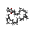
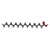


| #20: Chemical |  Mass: 55.845 Da / Num. of mol.: 2 / Source method: obtained synthetically / Formula: Fe Mass: 55.845 Da / Num. of mol.: 2 / Source method: obtained synthetically / Formula: Fe#21: Chemical | ChemComp-CL /  Mass: 35.453 Da / Num. of mol.: 4 / Source method: obtained synthetically / Formula: Cl Mass: 35.453 Da / Num. of mol.: 4 / Source method: obtained synthetically / Formula: Cl#22: Chemical | ChemComp-CLA /  Mass: 893.489 Da / Num. of mol.: 70 / Source method: obtained synthetically / Formula: C55H72MgN4O5 Mass: 893.489 Da / Num. of mol.: 70 / Source method: obtained synthetically / Formula: C55H72MgN4O5#23: Chemical | ChemComp-BCR /  Mass: 536.873 Da / Num. of mol.: 22 / Source method: obtained synthetically / Formula: C40H56 Mass: 536.873 Da / Num. of mol.: 22 / Source method: obtained synthetically / Formula: C40H56#24: Chemical | ChemComp-PL9 /  Mass: 749.201 Da / Num. of mol.: 4 / Source method: obtained synthetically / Formula: C53H80O2 / Feature type: SUBJECT OF INVESTIGATION Mass: 749.201 Da / Num. of mol.: 4 / Source method: obtained synthetically / Formula: C53H80O2 / Feature type: SUBJECT OF INVESTIGATION#25: Chemical | ChemComp-STE /  Mass: 284.477 Da / Num. of mol.: 42 / Source method: obtained synthetically / Formula: C18H36O2 Mass: 284.477 Da / Num. of mol.: 42 / Source method: obtained synthetically / Formula: C18H36O2#26: Chemical | ChemComp-LMG /  Mass: 787.158 Da / Num. of mol.: 14 / Source method: obtained synthetically / Formula: C45H86O10 Mass: 787.158 Da / Num. of mol.: 14 / Source method: obtained synthetically / Formula: C45H86O10#27: Chemical | ChemComp-SQD /  Mass: 795.116 Da / Num. of mol.: 8 / Source method: obtained synthetically / Formula: C41H78O12S Mass: 795.116 Da / Num. of mol.: 8 / Source method: obtained synthetically / Formula: C41H78O12S#29: Chemical |  Mass: 339.827 Da / Num. of mol.: 2 / Source method: obtained synthetically / Formula: CaMn4O5 / Feature type: SUBJECT OF INVESTIGATION Mass: 339.827 Da / Num. of mol.: 2 / Source method: obtained synthetically / Formula: CaMn4O5 / Feature type: SUBJECT OF INVESTIGATION#30: Chemical | ChemComp-PHO /  Mass: 871.200 Da / Num. of mol.: 4 / Source method: obtained synthetically / Formula: C55H74N4O5 Mass: 871.200 Da / Num. of mol.: 4 / Source method: obtained synthetically / Formula: C55H74N4O5#31: Chemical | ChemComp-LHG /  Mass: 722.970 Da / Num. of mol.: 10 / Source method: obtained synthetically / Formula: C38H75O10P / Comment: phospholipid*YM Mass: 722.970 Da / Num. of mol.: 10 / Source method: obtained synthetically / Formula: C38H75O10P / Comment: phospholipid*YM#33: Chemical |  Mass: 61.017 Da / Num. of mol.: 2 / Source method: obtained synthetically / Formula: CHO3 / Comment: pH buffer*YM Mass: 61.017 Da / Num. of mol.: 2 / Source method: obtained synthetically / Formula: CHO3 / Comment: pH buffer*YM#34: Chemical |  Mass: 616.487 Da / Num. of mol.: 2 / Source method: obtained synthetically / Formula: C34H32FeN4O4 Mass: 616.487 Da / Num. of mol.: 2 / Source method: obtained synthetically / Formula: C34H32FeN4O4#35: Chemical |  Mass: 618.503 Da / Num. of mol.: 2 / Source method: obtained synthetically / Formula: C34H34FeN4O4 Mass: 618.503 Da / Num. of mol.: 2 / Source method: obtained synthetically / Formula: C34H34FeN4O4#36: Water | ChemComp-HOH / | Mass: 18.015 Da / Num. of mol.: 2405 / Source method: isolated from a natural source / Formula: H2O |
|---|
-Details
| Has ligand of interest | Y |
|---|---|
| Has protein modification | Y |
-Experimental details
-Experiment
| Experiment | Method: ELECTRON MICROSCOPY |
|---|---|
| EM experiment | Aggregation state: PARTICLE / 3D reconstruction method: single particle reconstruction |
- Sample preparation
| Component | Name: dimeric core complex of PSII dPSII / Type: COMPLEX / Entity ID: #1-#19 / Source: NATURAL | |||||||||||||||||||||||||
|---|---|---|---|---|---|---|---|---|---|---|---|---|---|---|---|---|---|---|---|---|---|---|---|---|---|---|
| Molecular weight | Value: 750 kDa/nm / Experimental value: YES | |||||||||||||||||||||||||
| Source (natural) | Organism: Thermosynechococcus vestitus BP-1 (bacteria) | |||||||||||||||||||||||||
| Buffer solution | pH: 6.5 / Details: 100mM MES pH6.5, 5mM CaCl2, 5% glycerol, 0.03% DDM | |||||||||||||||||||||||||
| Buffer component |
| |||||||||||||||||||||||||
| Specimen | Conc.: 42.8 mg/ml / Embedding applied: NO / Shadowing applied: NO / Staining applied: NO / Vitrification applied: YES / Details: highly purified monodisperse sample of dPSIIcc | |||||||||||||||||||||||||
| Specimen support | Grid material: GOLD / Grid mesh size: 300 divisions/in. / Grid type: UltrAuFoil R1.2/1.3 | |||||||||||||||||||||||||
| Vitrification | Instrument: FEI VITROBOT MARK IV / Cryogen name: ETHANE / Humidity: 100 % / Chamber temperature: 277.15 K Details: The sample-on-grid was illuminated with one and two flashes of light |
- Electron microscopy imaging
| Experimental equipment |  Model: Titan Krios / Image courtesy: FEI Company |
|---|---|
| Microscopy | Model: FEI TITAN KRIOS |
| Electron gun | Electron source:  FIELD EMISSION GUN / Accelerating voltage: 300 kV / Illumination mode: FLOOD BEAM FIELD EMISSION GUN / Accelerating voltage: 300 kV / Illumination mode: FLOOD BEAM |
| Electron lens | Mode: BRIGHT FIELD / Nominal defocus max: 2000 nm / Nominal defocus min: 800 nm |
| Specimen holder | Cryogen: NITROGEN / Specimen holder model: FEI TITAN KRIOS AUTOGRID HOLDER |
| Image recording | Electron dose: 50 e/Å2 / Film or detector model: FEI FALCON IV (4k x 4k) |
- Processing
| EM software | Name: PHENIX / Version: 1.20.1_4487: / Category: model refinement | ||||||||||||||||||||||||
|---|---|---|---|---|---|---|---|---|---|---|---|---|---|---|---|---|---|---|---|---|---|---|---|---|---|
| CTF correction | Type: PHASE FLIPPING AND AMPLITUDE CORRECTION | ||||||||||||||||||||||||
| Symmetry | Point symmetry: C2 (2 fold cyclic) | ||||||||||||||||||||||||
| 3D reconstruction | Resolution: 1.71 Å / Resolution method: FSC 0.143 CUT-OFF / Num. of particles: 631270 / Symmetry type: POINT | ||||||||||||||||||||||||
| Atomic model building | Space: REAL | ||||||||||||||||||||||||
| Refine LS restraints |
|
