- EMDB-50019: cryoEM structure of Photosystem II averaged across S2-S3 states a... -
+
Open data
ID or keywords:
Loading...
-
Basic information
Entry
Database: EMDB / ID: EMD-50019
Title
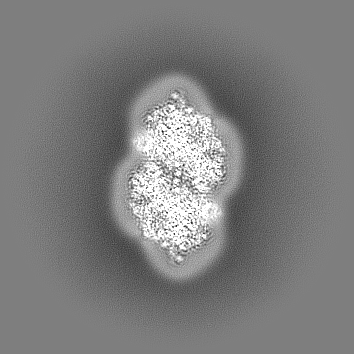
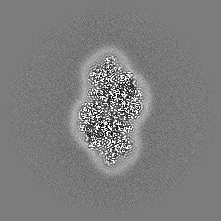
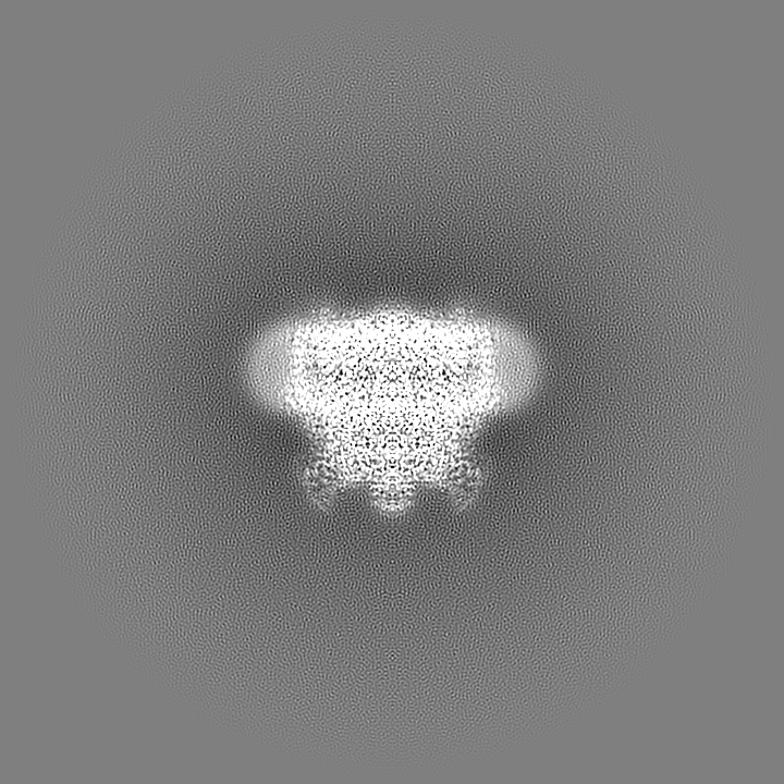
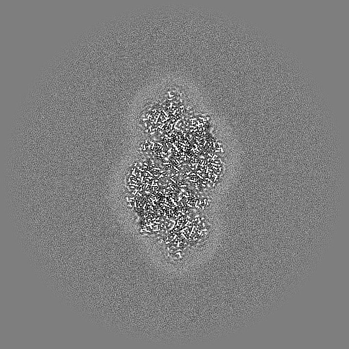
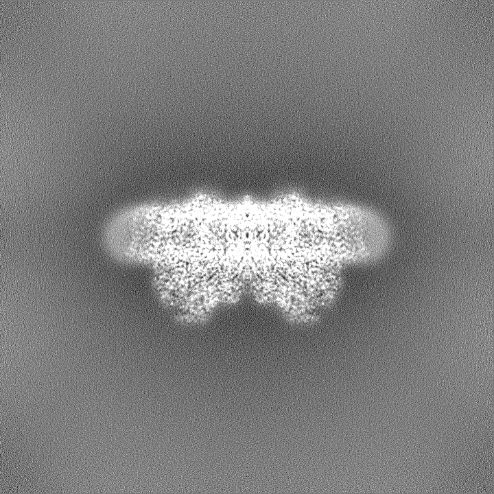
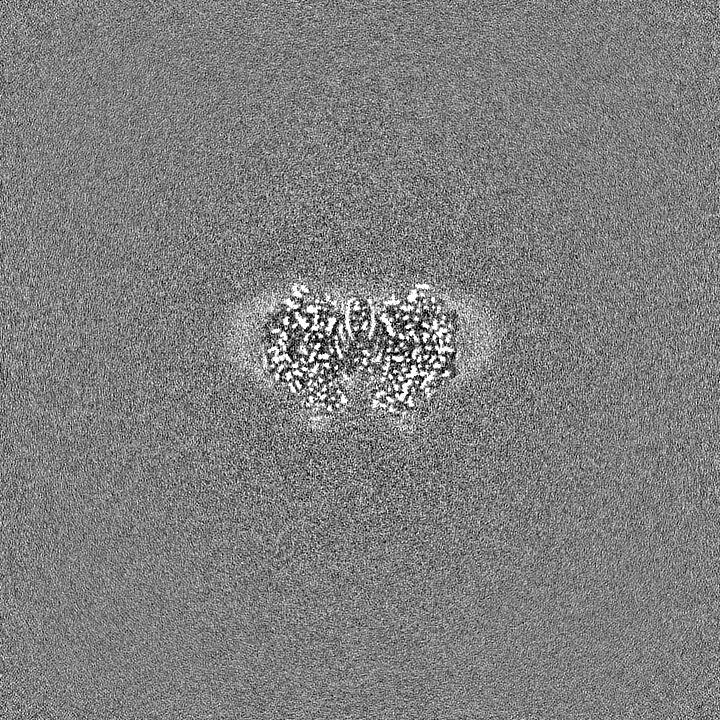
cryoEM structure of Photosystem II averaged across S2-S3 states at 1.71 Angstrom resolution
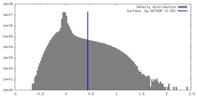
Map data
The original map before sharpening
Sample
Complex: dimeric core complex of PSII dPSII
Protein or peptide: x 19 types
Ligand: x 17 types
Keywords
Photosystem II core complex / Mn cluster / water oxidation / METAL BINDING PROTEIN
Function / homology
Function and homology information
photosystem II oxygen evolving complex / photosystem II assembly / oxygen evolving activity / photosystem II stabilization / photosystem II reaction center / photosystem II / photosynthetic electron transport chain / oxidoreductase activity, acting on diphenols and related substances as donors, oxygen as acceptor / response to herbicide / photosystem II ...photosystem II oxygen evolving complex / photosystem II assembly / oxygen evolving activity / photosystem II stabilization / photosystem II reaction center / photosystem II / photosynthetic electron transport chain / oxidoreductase activity, acting on diphenols and related substances as donors, oxygen as acceptor / response to herbicide / photosystem II / extrinsic component of membrane / plasma membrane-derived thylakoid membrane / photosynthetic electron transport in photosystem II / chlorophyll binding / photosynthesis, light reaction / phosphate ion binding / photosynthesis / respiratory electron transport chain / electron transfer activity / protein stabilization / iron ion binding / heme binding / metal ion binding Similarity search - Function
Photosystem II PsbU, oxygen evolving complex / Photosystem II 12 kDa extrinsic protein (PsbU) / Photosystem II PsbV, cytochrome c-550 precursor / Photosystem II cytochrome c-550 precursor / Cytochrome c-550 domain / Cytochrome c-550 domain / Photosystem II PsbX, type 1 subfamily / Photosystem II PsbJ / Photosystem II PsbJ superfamily / PsbJ ...Photosystem II PsbU, oxygen evolving complex / Photosystem II 12 kDa extrinsic protein (PsbU) / Photosystem II PsbV, cytochrome c-550 precursor / Photosystem II cytochrome c-550 precursor / Cytochrome c-550 domain / Cytochrome c-550 domain / Photosystem II PsbX, type 1 subfamily / Photosystem II PsbJ / Photosystem II PsbJ superfamily / PsbJ / Photosystem II PsbO, manganese-stabilising / Manganese-stabilising protein / photosystem II polypeptide / Photosystem II reaction centre protein Ycf12 / Photosystem II complex subunit Ycf12 / Photosystem II reaction centre M protein (PsbM) / Photosystem II PsbM superfamily / Photosystem II PsbM / Photosystem II PsbZ, reaction centre / Photosystem II PsbZ superfamily / YCF9 / Photosystem II PsbX / Photosystem II reaction centre X protein (PsbX) / Photosystem II PsbT / Photosystem II PsbL / Photosystem II CP43 reaction centre protein / Photosystem II PsbL superfamily / Photosystem II PsbT superfamily / Photosystem II CP43 reaction centre protein superfamily / Photosystem II reaction centre T protein / PsbL protein / Photosystem II PsbK / Photosystem II PsbK superfamily / Photosystem II 4 kDa reaction centre component / Photosystem II PsbI / Photosystem II CP47 reaction centre protein / Photosystem II PsbI superfamily / Photosystem II reaction centre I protein (PSII 4.8 kDa protein) / Photosystem II protein D1 / Photosystem II reaction centre protein H / Photosystem II D2 protein / Photosystem II cytochrome b559, conserved site / Photosystem II cytochrome b559, alpha subunit / Photosystem II cytochrome b559, beta subunit / Photosystem II cytochrome b559, N-terminal / Photosystem II cytochrome b559, alpha subunit, lumenal region / Photosystem II reaction centre protein H superfamily / Photosystem II cytochrome b559, alpha subunit superfamily / Cytochrome b559, alpha (gene psbE) and beta (gene psbF)subunits / Lumenal portion of Cytochrome b559, alpha (gene psbE) subunit / Photosystem II 10 kDa phosphoprotein / Cytochrome b559 subunits heme-binding site signature. / : / Photosystem antenna protein-like / Photosystem antenna protein-like superfamily / Photosystem II protein / Outer membrane protein/outer membrane enzyme PagP, beta-barrel / : / Photosynthetic reaction centre, L/M / Photosystem II protein D1/D2 superfamily / Photosynthetic reaction centre protein / Photosynthetic reaction center proteins signature. / Cytochrome c family profile. / Cytochrome c-like domain / Cytochrome c-like domain superfamily Similarity search - Domain/homology
Photosystem II extrinsic protein V / Photosystem II extrinsic protein O / Photosystem II protein D1 1 / Photosystem II reaction center protein J / Photosystem II D2 protein / Photosystem II reaction center protein M / Photosystem II reaction center protein Z / Photosystem II CP43 reaction center protein / Photosystem II reaction center protein L / Cytochrome b559 subunit beta ...Photosystem II extrinsic protein V / Photosystem II extrinsic protein O / Photosystem II protein D1 1 / Photosystem II reaction center protein J / Photosystem II D2 protein / Photosystem II reaction center protein M / Photosystem II reaction center protein Z / Photosystem II CP43 reaction center protein / Photosystem II reaction center protein L / Cytochrome b559 subunit beta / Cytochrome b559 subunit alpha / Photosystem II reaction center protein T / Photosystem II CP47 reaction center protein / Photosystem II reaction center protein H / Photosystem II reaction center protein Psb30 / Photosystem II reaction center protein I / Photosystem II reaction center protein K / Photosystem II extrinsic protein U / Photosystem II reaction center protein X Similarity search - Component
Biological species
Thermosynechococcus vestitus BP-1 (bacteria)
Method
single particle reconstruction / cryo EM / Resolution: 1.71 Å
Journal: Science / Year: 2024 Title: Cryo-electron microscopy reveals hydrogen positions and water networks in photosystem II. Authors: Rana Hussein / André Graça / Jack Forsman / A Orkun Aydin / Michael Hall / Julia Gaetcke / Petko Chernev / Petra Wendler / Holger Dobbek / Johannes Messinger / Athina Zouni / Wolfgang P Schröder / Abstract: Photosystem II starts the photosynthetic electron transport chain that converts solar energy into chemical energy and thus sustains life on Earth. It catalyzes two chemical reactions: water oxidation ...Photosystem II starts the photosynthetic electron transport chain that converts solar energy into chemical energy and thus sustains life on Earth. It catalyzes two chemical reactions: water oxidation to molecular oxygen and plastoquinone reduction. Coupling of electron and proton transfer is crucial for efficiency; however, the molecular basis of these processes remains speculative owing to uncertain water binding sites and the lack of experimentally determined hydrogen positions. We thus collected high-resolution cryo-electron microscopy data of fully hydrated photosystem II from the thermophilic cyanobacterium to a final resolution of 1.71 angstroms. The structure reveals several previously undetected partially occupied water binding sites and more than half of the hydrogen and proton positions. This clarifies the pathways of substrate water binding and plastoquinone B protonation.
Cryogen name: ETHANE / Chamber humidity: 100 % / Chamber temperature: 277.15 K / Instrument: FEI VITROBOT MARK IV Details: The sample-on-grid was illuminated with one and two flashes of light.
Details
highly purified monodisperse sample of dPSIIcc
-
Electron microscopy
Microscope
FEI TITAN KRIOS
Image recording
Film or detector model: FEI FALCON IV (4k x 4k) / Average electron dose: 50.0 e/Å2
Electron beam
Acceleration voltage: 300 kV / Electron source: FIELD EMISSION GUN
In the structure databanks used in Yorodumi, some data are registered as the other names, "COVID-19 virus" and "2019-nCoV". Here are the details of the virus and the list of structure data.
Jan 31, 2019. EMDB accession codes are about to change! (news from PDBe EMDB page)
EMDB accession codes are about to change! (news from PDBe EMDB page)
The allocation of 4 digits for EMDB accession codes will soon come to an end. Whilst these codes will remain in use, new EMDB accession codes will include an additional digit and will expand incrementally as the available range of codes is exhausted. The current 4-digit format prefixed with “EMD-” (i.e. EMD-XXXX) will advance to a 5-digit format (i.e. EMD-XXXXX), and so on. It is currently estimated that the 4-digit codes will be depleted around Spring 2019, at which point the 5-digit format will come into force.
The EM Navigator/Yorodumi systems omit the EMD- prefix.
Related info.:Q: What is EMD? / ID/Accession-code notation in Yorodumi/EM Navigator
Yorodumi is a browser for structure data from EMDB, PDB, SASBDB, etc.
This page is also the successor to EM Navigator detail page, and also detail information page/front-end page for Omokage search.
The word "yorodu" (or yorozu) is an old Japanese word meaning "ten thousand". "mi" (miru) is to see.
Related info.:EMDB / PDB / SASBDB / Comparison of 3 databanks / Yorodumi Search / Aug 31, 2016. New EM Navigator & Yorodumi / Yorodumi Papers / Jmol/JSmol / Function and homology information / Changes in new EM Navigator and Yorodumi
 Movie
Movie Controller

 Yorodumi
 Open data
 Basic information
 Map data
 Sample
 Keywords
 Function and homology information
 Thermosynechococcus vestitus BP-1 (bacteria)
 Authors
 Sweden,
  Germany, 3 items
  Citation
 Structure visualization
 Downloads & links
 emd_50019.png
 http://ftp.pdbj.org/pub/emdb/structures/EMD-50019


 Z (Sec.)
 Y (Row.)
 X (Col.)


 Sample components


 Processing
 Electron microscopy
 FIELD EMISSION GUN

