 Movie
Movie Controller

[English] 日本語
 Yorodumi
- PDB-9evr: Crystal structure of the kinetoplastid kinetochore protein KKT23 ... -
+ Open data

- Basic information
| Entry | Database: PDB / ID: 9evr | |||||||||
|---|---|---|---|---|---|---|---|---|---|---|
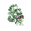
| Title | Crystal structure of the kinetoplastid kinetochore protein KKT23 N-terminal domain from Trypanosoma brucei | |||||||||
 Components Components | N-acetyltransferase domain-containing protein | |||||||||
 Keywords Keywords | CELL CYCLE / kinetochore / kinetoplastid / histone / acetyltransferase / mitosis | |||||||||
| Function / homology | acyltransferase activity, transferring groups other than amino-acyl groups / Acetyltransferase (GNAT) family / Gcn5-related N-acetyltransferase (GNAT) domain profile. / GNAT domain / Acyl-CoA N-acyltransferase / kinetochore / nucleus / cytoplasm / N-acetyltransferase domain-containing protein Function and homology information Function and homology information | |||||||||
| Biological species |  | |||||||||
| Method |  X-RAY DIFFRACTION / SYNCHROTRON / AB INITIO PHASING / Resolution: 2.7 Å X-RAY DIFFRACTION / SYNCHROTRON / AB INITIO PHASING / Resolution: 2.7 Å | |||||||||
 Authors Authors | Ludzia, P. / Ishii, M. / Akiyoshi, B. | |||||||||
| Funding support |  United Kingdom, United Kingdom,  Germany, 2items Germany, 2items
| |||||||||
 Citation Citation | Journal: Structure / Year: 2025 Title: The kinetoplastid kinetochore protein KKT23 acetyltransferase is a structural homolog of GCN5 that acetylates the histone H2A C-terminal tail. Authors: Ludzia, P. / Ishii, M. / Deak, G. / Spanos, C. / Wilson, M.D. / Redfield, C. / Akiyoshi, B. #1: Journal: Biorxiv / Year: 2024Title: The kinetoplastid kinetochore protein KKT23 acetyltransferase is a structural homolog of GCN5 that acetylates the histone H2A C-terminal tail Authors: Ludzia, P. / Ishii, M. / Akiyoshi, B. | |||||||||
| History |
|
- Structure visualization
| Structure viewer | Molecule: MolmilJmol/JSmol |
|---|
- Downloads & links
-Download
| PDBx/mmCIF format | 9evr.cif.gz | 33.3 KB | Display | PDBx/mmCIF format |
|---|---|---|---|---|
| PDB format | pdb9evr.ent.gz | 21.2 KB | Display | PDB format |
| PDBx/mmJSON format | 9evr.json.gz | Tree view | PDBx/mmJSON format | |
| Others |  Other downloads Other downloads |
-Validation report
| Summary document | 9evr_validation.pdf.gz | 424.9 KB | Display | wwPDB validaton report |
|---|---|---|---|---|
| Full document | 9evr_full_validation.pdf.gz | 425 KB | Display | |
| Data in XML | 9evr_validation.xml.gz | 3.8 KB | Display | |
| Data in CIF | 9evr_validation.cif.gz | 4.3 KB | Display | |
| Arichive directory | https://data.pdbj.org/pub/pdb/validation_reports/ev/9evrftp://data.pdbj.org/pub/pdb/validation_reports/ev/9evr | HTTPS FTP |
-Related structure data

-Links
 PDBj
PDBj


- Assembly
| Deposited unit | 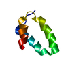
| ||||||||
|---|---|---|---|---|---|---|---|---|---|
| 1 |
| ||||||||
| Unit cell |
|
-Components
| #1: Protein | Mass: 8104.038 Da / Num. of mol.: 1 Source method: isolated from a genetically manipulated source Details: 6HIS-KKT23 2-70 Source: (gene. exp.) Gene: Tb10.70.0180 / Production host:  |
|---|---|
| Has protein modification | N |
-Experimental details
-Experiment
| Experiment | Method: X-RAY DIFFRACTION / Number of used crystals: 1 |
|---|
- Sample preparation
| Crystal | Density Matthews: 2.45 Å3/Da / Density % sol: 49.7 % |
|---|---|
| Crystal grow | Temperature: 277 K / Method: vapor diffusion, sitting drop / Details: 1M Sodium phosphate, pH 6.5, 12% w/v PEG 8000 |
-Data collection
| Diffraction | Mean temperature: 291 K / Serial crystal experiment: N | |||||||||||||||||||||
|---|---|---|---|---|---|---|---|---|---|---|---|---|---|---|---|---|---|---|---|---|---|---|
| Diffraction source | Source: SYNCHROTRON / Site: Diamond / Beamline: I04 / Wavelength: 0.9795 Å | |||||||||||||||||||||
| Detector | Type: DECTRIS EIGER X 16M / Detector: PIXEL / Date: Nov 28, 2021 | |||||||||||||||||||||
| Radiation | Protocol: SINGLE WAVELENGTH / Monochromatic (M) / Laue (L): M / Scattering type: x-ray | |||||||||||||||||||||
| Radiation wavelength | Wavelength: 0.9795 Å / Relative weight: 1 | |||||||||||||||||||||
| Reflection | Resolution: 2.49→42.73 Å / Num. obs: 3165 / % possible obs: 100 % / Redundancy: 18.4 % / CC1/2: 1 / Rrim(I) all: 0.106 / Net I/σ(I): 12.7 | |||||||||||||||||||||
| Reflection shell |
|
- Processing
| Software |
| ||||||||||||||||||||||||||||||||||||||||
|---|---|---|---|---|---|---|---|---|---|---|---|---|---|---|---|---|---|---|---|---|---|---|---|---|---|---|---|---|---|---|---|---|---|---|---|---|---|---|---|---|---|
| Refinement | Method to determine structure: AB INITIO PHASING / Resolution: 2.7→42.73 Å / SU ML: 0.17 / Cross valid method: FREE R-VALUE / σ(F): 2.15 / Phase error: 24.33 / Stereochemistry target values: ML
| ||||||||||||||||||||||||||||||||||||||||
| Solvent computation | Shrinkage radii: 0.9 Å / VDW probe radii: 1.11 Å / Solvent model: FLAT BULK SOLVENT MODEL | ||||||||||||||||||||||||||||||||||||||||
| Refinement step | Cycle: LAST / Resolution: 2.7→42.73 Å
| ||||||||||||||||||||||||||||||||||||||||
| Refine LS restraints |
| ||||||||||||||||||||||||||||||||||||||||
| LS refinement shell | Resolution: 2.7→42.73 Å
| ||||||||||||||||||||||||||||||||||||||||
| Refinement TLS params. | Method: refined / Origin x: -1.1699 Å / Origin y: -30.0489 Å / Origin z: 21.1297 Å
| ||||||||||||||||||||||||||||||||||||||||
| Refinement TLS group | Selection details: (chain 'A' and resid 17 through 64) |
