 Movie
Movie Controller


+ Open data

- Basic information
| Entry | Database: PDB / ID: 9et3 | ||||||||||||
|---|---|---|---|---|---|---|---|---|---|---|---|---|---|
| Title | CDK2-cyclin A in complex with FragLite 10 | ||||||||||||
 Components Components |
| ||||||||||||
 Keywords Keywords | CELL CYCLE / cyclin-dependent kinase / FragLite / CDK2 / cyclin A / Fragment | ||||||||||||
| Function / homology |  Function and homology information Function and homology informationcell cycle G1/S phase transition / cyclin-dependent protein serine/threonine kinase regulator activity / cyclin A1-CDK2 complex / cyclin E2-CDK2 complex / cyclin E1-CDK2 complex / cyclin A2-CDK2 complex / positive regulation of DNA-templated DNA replication initiation / G2 Phase / Y chromosome / cyclin-dependent protein kinase activity ...cell cycle G1/S phase transition / cyclin-dependent protein serine/threonine kinase regulator activity / cyclin A1-CDK2 complex / cyclin E2-CDK2 complex / cyclin E1-CDK2 complex / cyclin A2-CDK2 complex / positive regulation of DNA-templated DNA replication initiation / G2 Phase / Y chromosome / cyclin-dependent protein kinase activity / regulation of heterochromatin organization / Phosphorylation of proteins involved in G1/S transition by active Cyclin E:Cdk2 complexes / positive regulation of heterochromatin formation / p53-Dependent G1 DNA Damage Response / X chromosome / PTK6 Regulates Cell Cycle / regulation of anaphase-promoting complex-dependent catabolic process / Defective binding of RB1 mutants to E2F1,(E2F2, E2F3) / centriole replication / Regulation of APC/C activators between G1/S and early anaphase / telomere maintenance in response to DNA damage / regulation of DNA replication / centrosome duplication / microtubule organizing center / G0 and Early G1 / Telomere Extension By Telomerase / Activation of the pre-replicative complex / cyclin-dependent kinase / cyclin-dependent protein serine/threonine kinase activity / TP53 Regulates Transcription of Genes Involved in G1 Cell Cycle Arrest / Activation of ATR in response to replication stress / Regulation of MITF-M-dependent genes involved in cell cycle and proliferation / Cajal body / Cyclin E associated events during G1/S transition / Cyclin A:Cdk2-associated events at S phase entry / cyclin-dependent protein kinase holoenzyme complex / Cyclin A/B1/B2 associated events during G2/M transition / regulation of G2/M transition of mitotic cell cycle / condensed chromosome / mitotic G1 DNA damage checkpoint signaling / negative regulation of protein localization to chromatin / cellular response to nitric oxide / post-translational protein modification / regulation of mitotic cell cycle / cyclin binding / positive regulation of DNA replication / peptidyl-serine phosphorylation / male germ cell nucleus / meiotic cell cycle / potassium ion transport / G1/S transition of mitotic cell cycle / DNA Damage/Telomere Stress Induced Senescence / Meiotic recombination / G2/M transition of mitotic cell cycle / CDK-mediated phosphorylation and removal of Cdc6 / Transcriptional regulation of granulopoiesis / SCF(Skp2)-mediated degradation of p27/p21 / Orc1 removal from chromatin / cellular senescence / Cyclin D associated events in G1 / Regulation of TP53 Degradation / nuclear envelope / Factors involved in megakaryocyte development and platelet production / regulation of gene expression / Processing of DNA double-strand break ends / Senescence-Associated Secretory Phenotype (SASP) / transcription regulator complex / Regulation of TP53 Activity through Phosphorylation / protein phosphorylation / Ras protein signal transduction / DNA replication / chromosome, telomeric region / endosome / chromatin remodeling / protein domain specific binding / protein serine kinase activity / cell division / DNA repair / protein serine/threonine kinase activity / positive regulation of cell population proliferation / centrosome / protein kinase binding / positive regulation of DNA-templated transcription / DNA-templated transcription / magnesium ion binding / negative regulation of transcription by RNA polymerase II / signal transduction / nucleoplasm / ATP binding / nucleus / cytoplasm / cytosol Similarity search - Function | ||||||||||||
| Biological species |   Homo sapiens (human) Homo sapiens (human) | ||||||||||||
| Method |  X-RAY DIFFRACTION / SYNCHROTRON / MOLECULAR REPLACEMENT / Resolution: 2.34 Å X-RAY DIFFRACTION / SYNCHROTRON / MOLECULAR REPLACEMENT / Resolution: 2.34 Å | ||||||||||||
 Authors Authors | Hope, I. / Martin, M.P. / Waring, M.J. / Noble, M.E.M. / Endicott, J.A. / Tatum, N.J. | ||||||||||||
| Funding support |  United Kingdom, 3items United Kingdom, 3items
| ||||||||||||
 Citation Citation | Journal: Structure / Year: 2025 Title: Crystallographic fragment screening of CDK2-cyclin A: FragLites map sites of protein-protein interaction. Authors: Hope, I. / Martin, M.P. / Jiang, Z. / Waring, M.J. / Noble, M.E.M. / Endicott, J.A. / Tatum, N.J. | ||||||||||||
| History |
|
- Structure visualization
| Structure viewer | Molecule: MolmilJmol/JSmol |
|---|
- Downloads & links
-Download
| PDBx/mmCIF format | 9et3.cif.gz | 240.3 KB | Display | PDBx/mmCIF format |
|---|---|---|---|---|
| PDB format | pdb9et3.ent.gz | 190.2 KB | Display | PDB format |
| PDBx/mmJSON format | 9et3.json.gz | Tree view | PDBx/mmJSON format | |
| Others |  Other downloads Other downloads |
-Validation report
| Arichive directory | https://data.pdbj.org/pub/pdb/validation_reports/et/9et3ftp://data.pdbj.org/pub/pdb/validation_reports/et/9et3 | HTTPS FTP |
|---|
-Related structure data
| Related structure data | 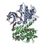 9esjC  9eskC  9eslC 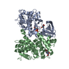 9esnC  9esoC  9espC  9esqC  9esrC 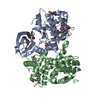 9essC  9esuC  9esvC  9eswC  9esxC  9esyC  9eszC  9et0C  9et1C  9et2C  9et4C  9et5C  9et6C  9et7C  9et8C  9et9C  9etaC  9etbC  9etpC C: citing same article ( |
|---|---|
| Similar structure data |
-Links
 PDBj
PDBj


- Assembly
| Deposited unit | 
| ||||||||
|---|---|---|---|---|---|---|---|---|---|
| 1 | 
| ||||||||
| 2 | 
| ||||||||
| Unit cell |
|
-Components
| #1: Protein | Mass: 30817.473 Da / Num. of mol.: 2 Source method: isolated from a genetically manipulated source Source: (gene. exp.)  #2: Protein | Mass: 34354.770 Da / Num. of mol.: 2 Source method: isolated from a genetically manipulated source Source: (gene. exp.) Homo sapiens (human) / Gene: CDK2, CDKN2 / Plasmid: PGEX6P1 / Production host: #3: Chemical | ChemComp-1P8 /   Mass: 212.043 Da / Num. of mol.: 14 / Source method: isolated from a natural source / Formula: C8H6BrNO / Feature type: SUBJECT OF INVESTIGATION Mass: 212.043 Da / Num. of mol.: 14 / Source method: isolated from a natural source / Formula: C8H6BrNO / Feature type: SUBJECT OF INVESTIGATION#4: Water | ChemComp-HOH / |  Mass: 18.015 Da / Num. of mol.: 298 / Source method: isolated from a natural source / Formula: H2O Mass: 18.015 Da / Num. of mol.: 298 / Source method: isolated from a natural source / Formula: H2OHas ligand of interest | Y | Has protein modification | Y | |
|---|
-Experimental details
-Experiment
| Experiment | Method: X-RAY DIFFRACTION / Number of used crystals: 1 |
|---|
- Sample preparation
| Crystal | Density Matthews: 2.97 Å3/Da / Density % sol: 58.56 % |
|---|---|
| Crystal grow | Temperature: 277 K / Method: vapor diffusion, sitting drop / pH: 7 Details: Protein at 10 mg/ml. 0.6 to 0.8 M KCl, 0.9 to 1.2 M (NH4)2SO4, and 100 mM HEPES pH 7.0 PH range: 7-7.5 |
-Data collection
| Diffraction | Mean temperature: 100 K / Serial crystal experiment: N |
|---|---|
| Diffraction source | Source: SYNCHROTRON / Site: Diamond / Beamline: I03 / Wavelength: 0.91839 Å |
| Detector | Type: DECTRIS PILATUS 6M / Detector: PIXEL / Date: May 16, 2020 |
| Radiation | Protocol: SINGLE WAVELENGTH / Monochromatic (M) / Laue (L): M / Scattering type: x-ray |
| Radiation wavelength | Wavelength: 0.91839 Å / Relative weight: 1 |
| Reflection | Resolution: 2.34→148.04 Å / Num. obs: 59478 / % possible obs: 94.4 % / Redundancy: 13.5 % / CC1/2: 0.999 / Rmerge(I) obs: 0.116 / Net I/σ(I): 16.8 |
| Reflection shell | Resolution: 2.34→2.4 Å / Redundancy: 14.1 % / Rmerge(I) obs: 1.624 / Mean I/σ(I) obs: 1.9 / Num. unique obs: 4870 / CC1/2: 0.658 / % possible all: 100 |
- Processing
| Software |
| |||||||||||||||||||||||||||||||||||||||||||||||||||||||||||||||||||||||||||||||||||||||||||||||||||||||||||||||||||||||||||||||||||||||||||||||||||||||||||||||||||||||||||||||||||||||||||||||||||||||||||||||||||||||||||||||||||||||
|---|---|---|---|---|---|---|---|---|---|---|---|---|---|---|---|---|---|---|---|---|---|---|---|---|---|---|---|---|---|---|---|---|---|---|---|---|---|---|---|---|---|---|---|---|---|---|---|---|---|---|---|---|---|---|---|---|---|---|---|---|---|---|---|---|---|---|---|---|---|---|---|---|---|---|---|---|---|---|---|---|---|---|---|---|---|---|---|---|---|---|---|---|---|---|---|---|---|---|---|---|---|---|---|---|---|---|---|---|---|---|---|---|---|---|---|---|---|---|---|---|---|---|---|---|---|---|---|---|---|---|---|---|---|---|---|---|---|---|---|---|---|---|---|---|---|---|---|---|---|---|---|---|---|---|---|---|---|---|---|---|---|---|---|---|---|---|---|---|---|---|---|---|---|---|---|---|---|---|---|---|---|---|---|---|---|---|---|---|---|---|---|---|---|---|---|---|---|---|---|---|---|---|---|---|---|---|---|---|---|---|---|---|---|---|---|---|---|---|---|---|---|---|---|---|---|---|---|---|---|---|---|---|
| Refinement | Method to determine structure: MOLECULAR REPLACEMENT / Resolution: 2.34→99.483 Å / Cor.coef. Fo:Fc: 0.964 / Cor.coef. Fo:Fc free: 0.948 / WRfactor Rfree: 0.207 / WRfactor Rwork: 0.17 / Average fsc free: 0.9573 / Average fsc work: 0.9706 / Cross valid method: THROUGHOUT / ESU R: 0.305 / ESU R Free: 0.217 / Details: Hydrogens have not been used
| |||||||||||||||||||||||||||||||||||||||||||||||||||||||||||||||||||||||||||||||||||||||||||||||||||||||||||||||||||||||||||||||||||||||||||||||||||||||||||||||||||||||||||||||||||||||||||||||||||||||||||||||||||||||||||||||||||||||
| Solvent computation | Ion probe radii: 0.8 Å / Shrinkage radii: 0.8 Å / VDW probe radii: 1.2 Å / Solvent model: MASK BULK SOLVENT | |||||||||||||||||||||||||||||||||||||||||||||||||||||||||||||||||||||||||||||||||||||||||||||||||||||||||||||||||||||||||||||||||||||||||||||||||||||||||||||||||||||||||||||||||||||||||||||||||||||||||||||||||||||||||||||||||||||||
| Displacement parameters | Biso mean: 58.129 Å2
| |||||||||||||||||||||||||||||||||||||||||||||||||||||||||||||||||||||||||||||||||||||||||||||||||||||||||||||||||||||||||||||||||||||||||||||||||||||||||||||||||||||||||||||||||||||||||||||||||||||||||||||||||||||||||||||||||||||||
| Refinement step | Cycle: LAST / Resolution: 2.34→99.483 Å
| |||||||||||||||||||||||||||||||||||||||||||||||||||||||||||||||||||||||||||||||||||||||||||||||||||||||||||||||||||||||||||||||||||||||||||||||||||||||||||||||||||||||||||||||||||||||||||||||||||||||||||||||||||||||||||||||||||||||
| Refine LS restraints |
| |||||||||||||||||||||||||||||||||||||||||||||||||||||||||||||||||||||||||||||||||||||||||||||||||||||||||||||||||||||||||||||||||||||||||||||||||||||||||||||||||||||||||||||||||||||||||||||||||||||||||||||||||||||||||||||||||||||||
| LS refinement shell | Refine-ID: X-RAY DIFFRACTION / Total num. of bins used: 20
|
