+ Open data
- Basic information
| Entry | Database: PDB / ID: 9em9 | |||||||||||||||||||||||||||||||||||||||
|---|---|---|---|---|---|---|---|---|---|---|---|---|---|---|---|---|---|---|---|---|---|---|---|---|---|---|---|---|---|---|---|---|---|---|---|---|---|---|---|---|
| Title | Structure of SynDLP MGD with GMPPNP | |||||||||||||||||||||||||||||||||||||||
 Components Components | Slr0869 protein | |||||||||||||||||||||||||||||||||||||||
 Keywords Keywords | LIPID BINDING PROTEIN / BDLP / cyanobacteria / membrane remodeling / GTPase | |||||||||||||||||||||||||||||||||||||||
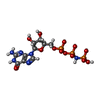
| Function / homology | Mitofusin family / Dynamin, N-terminal / Dynamin family / GTPase activity / GTP binding / P-loop containing nucleoside triphosphate hydrolase / membrane / PHOSPHOAMINOPHOSPHONIC ACID-GUANYLATE ESTER / Slr0869 protein Function and homology information Function and homology information | |||||||||||||||||||||||||||||||||||||||
| Biological species |  | |||||||||||||||||||||||||||||||||||||||
| Method | ELECTRON MICROSCOPY / single particle reconstruction / cryo EM / Resolution: 3.76 Å | |||||||||||||||||||||||||||||||||||||||
 Authors Authors | Junglas, B. / Gewehr, L. / Schoennenbeck, P. / Schneider, D. / Sachse, C. | |||||||||||||||||||||||||||||||||||||||
| Funding support | European Union, 1items
| |||||||||||||||||||||||||||||||||||||||
 Citation Citation |  Journal: Cell Rep / Year: 2024 Journal: Cell Rep / Year: 2024Title: Structural basis for GTPase activity and conformational changes of the bacterial dynamin-like protein SynDLP. Authors: Benedikt Junglas / Lucas Gewehr / Lara Mernberger / Philipp Schönnenbeck / Ruven Jilly / Nadja Hellmann / Dirk Schneider / Carsten Sachse /  Abstract: SynDLP, a dynamin-like protein (DLP) encoded in the cyanobacterium Synechocystis sp. PCC 6803, has recently been identified to be structurally highly similar to eukaryotic dynamins. To elucidate ...SynDLP, a dynamin-like protein (DLP) encoded in the cyanobacterium Synechocystis sp. PCC 6803, has recently been identified to be structurally highly similar to eukaryotic dynamins. To elucidate structural changes during guanosine triphosphate (GTP) hydrolysis, we solved the cryoelectron microscopy (cryo-EM) structures of oligomeric full-length SynDLP after addition of guanosine diphosphate (GDP) at 4.1 Å and GTP at 3.6-Å resolution as well as a GMPPNP-bound dimer structure of a minimal G-domain construct of SynDLP at 3.8-Å resolution. In comparison with what has been seen in the previously resolved apo structure, we found that the G-domain is tilted upward relative to the stalk upon GTP hydrolysis and that the G-domain dimerizes via an additional extended dimerization domain not present in canonical G-domains. When incubated with lipid vesicles, we observed formation of irregular tubular SynDLP assemblies that interact with negatively charged lipids. Here, we provide the structural framework of a series of different functional SynDLP assembly states during GTP turnover. | |||||||||||||||||||||||||||||||||||||||
| History |
|
- Structure visualization
| Structure viewer | Molecule:  Molmil Molmil Jmol/JSmol Jmol/JSmol |
|---|
- Downloads & links
- Download
| PDBx/mmCIF format |  9em9.cif.gz 9em9.cif.gz | 186.9 KB | Display |  PDBx/mmCIF format PDBx/mmCIF format |
|---|---|---|---|---|
| PDB format |  pdb9em9.ent.gz pdb9em9.ent.gz | 148.9 KB | Display |  PDB format PDB format |
| PDBx/mmJSON format |  9em9.json.gz 9em9.json.gz | Tree view |  PDBx/mmJSON format PDBx/mmJSON format | |
| Others |  Other downloads Other downloads |
-Validation report
| Arichive directory |  https://data.pdbj.org/pub/pdb/validation_reports/em/9em9 https://data.pdbj.org/pub/pdb/validation_reports/em/9em9 ftp://data.pdbj.org/pub/pdb/validation_reports/em/9em9 ftp://data.pdbj.org/pub/pdb/validation_reports/em/9em9 | HTTPS FTP |
|---|
-Related structure data
| Related structure data |  19814MC  9em7C  9em8C M: map data used to model this data C: citing same article ( |
|---|---|
| Similar structure data | Similarity search - Function & homology  F&H Search F&H Search |
- Links
Links
- Assembly
| Deposited unit | 
|
|---|---|
| 1 |
|
- Components
| #1: Protein | Mass: 59291.410 Da / Num. of mol.: 2 Mutation: GS Linker replaced BSE Domain,truncated Stalk Domain Source method: isolated from a genetically manipulated source Source: (gene. exp.)   #2: Chemical | #3: Chemical | Has ligand of interest | Y | Has protein modification | Y | |
|---|
-Experimental details
-Experiment
| Experiment | Method: ELECTRON MICROSCOPY |
|---|---|
| EM experiment | Aggregation state: PARTICLE / 3D reconstruction method: single particle reconstruction |
- Sample preparation
| Component | Name: SynDLP MGD / Type: COMPLEX / Entity ID: #1 / Source: RECOMBINANT |
|---|---|
| Molecular weight | Experimental value: NO |
| Source (natural) | Organism:  |
| Source (recombinant) | Organism:  |
| Buffer solution | pH: 7.5 |
| Specimen | Embedding applied: NO / Shadowing applied: NO / Staining applied: NO / Vitrification applied: YES |
| Vitrification | Cryogen name: ETHANE |
- Electron microscopy imaging
| Experimental equipment |  Model: Talos Arctica / Image courtesy: FEI Company |
|---|---|
| Microscopy | Model: FEI TALOS ARCTICA |
| Electron gun | Electron source:  FIELD EMISSION GUN / Accelerating voltage: 200 kV / Illumination mode: FLOOD BEAM FIELD EMISSION GUN / Accelerating voltage: 200 kV / Illumination mode: FLOOD BEAM |
| Electron lens | Mode: BRIGHT FIELD / Nominal defocus max: 3500 nm / Nominal defocus min: 1000 nm |
| Image recording | Electron dose: 44.5 e/Å2 / Film or detector model: GATAN K3 BIOQUANTUM (6k x 4k) |
- Processing
| EM software | Name: PHENIX / Category: model refinement | ||||||||||||||||||||||||
|---|---|---|---|---|---|---|---|---|---|---|---|---|---|---|---|---|---|---|---|---|---|---|---|---|---|
| CTF correction | Type: PHASE FLIPPING AND AMPLITUDE CORRECTION | ||||||||||||||||||||||||
| 3D reconstruction | Resolution: 3.76 Å / Resolution method: FSC 0.143 CUT-OFF / Num. of particles: 219630 / Symmetry type: POINT | ||||||||||||||||||||||||
| Refine LS restraints |
|
 Movie
Movie Controller


 PDBj
PDBj


