Movie
Movie Controller


+ Open data

- Basic information
| Entry | Database: PDB / ID: 9ek3 | ||||||||||||
|---|---|---|---|---|---|---|---|---|---|---|---|---|---|
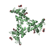
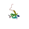
| Title | HIV-1 immature WT matrix protein p17 lattice | ||||||||||||
 Components Components | Matrix protein p17 | ||||||||||||
 Keywords Keywords | VIRAL PROTEIN / MATRIX / HIV-1 / p17 / HIV-1 p17 / VIRUS / GAG / STRUCTURAL PROTEIN | ||||||||||||
| Function / homology |  Function and homology information Function and homology informationHIV-1 retropepsin / symbiont-mediated activation of host apoptosis / retroviral ribonuclease H / exoribonuclease H / exoribonuclease H activity / host multivesicular body / DNA integration / viral genome integration into host DNA / RNA-directed DNA polymerase / establishment of integrated proviral latency ...HIV-1 retropepsin / symbiont-mediated activation of host apoptosis / retroviral ribonuclease H / exoribonuclease H / exoribonuclease H activity / host multivesicular body / DNA integration / viral genome integration into host DNA / RNA-directed DNA polymerase / establishment of integrated proviral latency / viral penetration into host nucleus / RNA stem-loop binding / RNA-directed DNA polymerase activity / RNA-DNA hybrid ribonuclease activity / Transferases; Transferring phosphorus-containing groups; Nucleotidyltransferases / host cell / viral nucleocapsid / DNA recombination / DNA-directed DNA polymerase / aspartic-type endopeptidase activity / Hydrolases; Acting on ester bonds / DNA-directed DNA polymerase activity / symbiont-mediated suppression of host gene expression / viral translational frameshifting / lipid binding / symbiont entry into host cell / host cell nucleus / host cell plasma membrane / virion membrane / structural molecule activity / proteolysis / DNA binding / zinc ion binding / membrane Similarity search - Function | ||||||||||||
| Biological species |   Human immunodeficiency virus type 1 Human immunodeficiency virus type 1 | ||||||||||||
| Method | ELECTRON MICROSCOPY / subtomogram averaging / cryo EM / Resolution: 8 Å | ||||||||||||
 Authors Authors | Rey, J.S. / Perilla, J.R. / Chen, L. / Zhang, P. | ||||||||||||
| Funding support |  United States, 3items United States, 3items
| ||||||||||||
 Citation Citation | Journal: Sci Adv / Year: 2025 Title: Structural maturation of the matrix lattice is not required for HIV-1 particle infectivity. Authors: Long Chen / Yuta Hikichi / Juan S Rey / Caner Akıl / Yanan Zhu / Hana Veler / Yao Shen / Juan R Perilla / Eric O Freed / Peijun Zhang /   Abstract: During HIV-1 maturation, the matrix (MA) lattice underlying the viral membrane undergoes a structural rearrangement, and the newly released capsid (CA) protein forms a mature CA. While it is well ...During HIV-1 maturation, the matrix (MA) lattice underlying the viral membrane undergoes a structural rearrangement, and the newly released capsid (CA) protein forms a mature CA. While it is well established that CA formation is essential for particle infectivity, the functional role of MA structural maturation remains unclear. Here, we examine maturation of an MA triple mutant, L20K/E73K/A82T, which, despite replicating similarly to wild-type (WT) in some cell lines, exhibits distinct biochemical behaviors that suggest altered MA-MA interactions. Cryo-electron tomography with subtomogram averaging reveals that, although the MA lattice in immature L20K/E73K/A82T virions closely resembles that of the WT, mature L20K/E73K/A82T virions lack a detectable MA lattice. All-atom molecular dynamics simulations suggest that this absence results from destabilized inter-trimer MA interactions in mature L20K/E73K/A82T mutant virions. These findings suggest that an ordered, membrane-associated mature MA lattice is not essential for HIV-1 infectivity, providing insights into the structural requirements for HIV-1 particle maturation and generation of infectious particles. | ||||||||||||
| History |
|
- Structure visualization
| Structure viewer | Molecule: MolmilJmol/JSmol |
|---|
- Downloads & links
-Download
| PDBx/mmCIF format | 9ek3.cif.gz | 746.5 KB | Display | PDBx/mmCIF format |
|---|---|---|---|---|
| PDB format | pdb9ek3.ent.gz | 614.7 KB | Display | PDB format |
| PDBx/mmJSON format | 9ek3.json.gz | Tree view | PDBx/mmJSON format | |
| Others |  Other downloads Other downloads |
-Validation report
| Summary document | 9ek3_validation.pdf.gz | 423.8 KB | Display | wwPDB validaton report |
|---|---|---|---|---|
| Full document | 9ek3_full_validation.pdf.gz | 434.2 KB | Display | |
| Data in XML | 9ek3_validation.xml.gz | 69.6 KB | Display | |
| Data in CIF | 9ek3_validation.cif.gz | 117.6 KB | Display | |
| Arichive directory | https://data.pdbj.org/pub/pdb/validation_reports/ek/9ek3ftp://data.pdbj.org/pub/pdb/validation_reports/ek/9ek3 | HTTPS FTP |
-Related structure data
| Related structure data |  9ek1C  9ek2C M: map data used to model this data C: citing same article ( |
|---|---|
| Similar structure data |


-Links
 PDBj
PDBj


- Assembly
| Deposited unit | 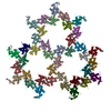
|
|---|---|
| 1 |
|
-Components
| #1: Protein | Mass: 13084.983 Da / Num. of mol.: 39 Source method: isolated from a genetically manipulated source Source: (gene. exp.) Human immunodeficiency virus type 1 / Strain: pNL4-3 / Gene: gag-pol / Production host:  Homo sapiens (human) / References: UniProt: P12497 Homo sapiens (human) / References: UniProt: P12497#2: Chemical | ChemComp-MYR /   Mass: 228.371 Da / Num. of mol.: 39 / Source method: obtained synthetically / Formula: C14H28O2 Mass: 228.371 Da / Num. of mol.: 39 / Source method: obtained synthetically / Formula: C14H28O2Has ligand of interest | N | Has protein modification | Y | |
|---|
-Experimental details
-Experiment
| Experiment | Method: ELECTRON MICROSCOPY |
|---|---|
| EM experiment | Aggregation state: PARTICLE / 3D reconstruction method: subtomogram averaging |
- Sample preparation
| Component | Name: HIV-1 / Type: VIRUS / Entity ID: #1 / Source: RECOMBINANT |
|---|---|
| Source (natural) | Organism: HIV-1 (virus) / Strain: pNL4-3 |
| Source (recombinant) | Organism: Homo sapiens (human) |
| Details of virus | Empty: NO / Enveloped: YES / Isolate: STRAIN / Type: VIRUS-LIKE PARTICLE |
| Buffer solution | pH: 7.4 |
| Specimen | Embedding applied: NO / Shadowing applied: NO / Staining applied: NO / Vitrification applied: YES |
| Vitrification | Cryogen name: ETHANE |
- Electron microscopy imaging
| Experimental equipment |  Model: Titan Krios / Image courtesy: FEI Company |
|---|---|
| Microscopy | Model: TFS KRIOS |
| Electron gun | Electron source:  FIELD EMISSION GUN / Accelerating voltage: 300 kV / Illumination mode: FLOOD BEAM FIELD EMISSION GUN / Accelerating voltage: 300 kV / Illumination mode: FLOOD BEAM |
| Electron lens | Mode: BRIGHT FIELD / Nominal defocus max: 5500 nm / Nominal defocus min: 1500 nm |
| Image recording | Electron dose: 3 e/Å2 / Avg electron dose per subtomogram: 123 e/Å2 / Film or detector model: FEI FALCON IV (4k x 4k) |
- Processing
| EM software |
| |||||||||||||||||||||
|---|---|---|---|---|---|---|---|---|---|---|---|---|---|---|---|---|---|---|---|---|---|---|
| CTF correction | Type: PHASE FLIPPING AND AMPLITUDE CORRECTION | |||||||||||||||||||||
| Symmetry | Point symmetry: C3 (3 fold cyclic) | |||||||||||||||||||||
| 3D reconstruction | Resolution: 8 Å / Resolution method: FSC 0.143 CUT-OFF / Num. of particles: 18262 / Symmetry type: POINT | |||||||||||||||||||||
| EM volume selection | Num. of tomograms: 89 / Num. of volumes extracted: 18262 | |||||||||||||||||||||
| Atomic model building |
|