 Movie
Movie Controller

[English] 日本語
 Yorodumi
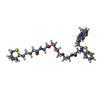
- PDB-9ehb: Structure of short Lettuce aptamer (C14T variant) bound with TO1-... -
+ Open data

- Basic information
| Entry | Database: PDB / ID: 9ehb | ||||||
|---|---|---|---|---|---|---|---|
| Title | Structure of short Lettuce aptamer (C14T variant) bound with TO1-biotin. | ||||||
 Components Components | DNA 53-mer | ||||||
 Keywords Keywords | DNA / Structure of short Lettuce aptamer (C14T variant) bound with TO1-biotin. | ||||||
| Function / homology | DIMETHYLFORMAMIDE / Chem-HZD / : / DNA / DNA (> 10) Function and homology information Function and homology information | ||||||
| Biological species | synthetic construct (others) | ||||||
| Method |  X-RAY DIFFRACTION / SYNCHROTRON / MOLECULAR REPLACEMENT / Resolution: 2.03 Å X-RAY DIFFRACTION / SYNCHROTRON / MOLECULAR REPLACEMENT / Resolution: 2.03 Å | ||||||
 Authors Authors | Koomullam, N. / Batchelder-Schwab, E. / Mao, C. | ||||||
| Funding support |  United States, 1items United States, 1items
| ||||||
 Citation Citation | Journal: ACS Sens / Year: 2026 Title: Fluorogenic Aptamer Optimization on a Massively Parallel Sequencing Platform. Authors: Kuo, Y.A. / Chen, Y.I. / Siraj, N. / He, Y. / Yang, Z. / Wang, Y. / Batchelder-Schwab, E.J. / Korkmaz, Z. / Yonas, S. / Nguyen, T.D. / Hong, S. / Nguyen, A.T. / Kim, S. / Seifi, S. / Fan, P. ...Authors: Kuo, Y.A. / Chen, Y.I. / Siraj, N. / He, Y. / Yang, Z. / Wang, Y. / Batchelder-Schwab, E.J. / Korkmaz, Z. / Yonas, S. / Nguyen, T.D. / Hong, S. / Nguyen, A.T. / Kim, S. / Seifi, S. / Fan, P.H. / Wu, Y. / Liu, H.W. / Lu, Y. / Ren, P. / Mao, C. / Yeh, H.C. | ||||||
| History |
|
- Structure visualization
| Structure viewer | Molecule: MolmilJmol/JSmol |
|---|
- Downloads & links
-Download
| PDBx/mmCIF format | 9ehb.cif.gz | 87.8 KB | Display | PDBx/mmCIF format |
|---|---|---|---|---|
| PDB format | pdb9ehb.ent.gz | 55.2 KB | Display | PDB format |
| PDBx/mmJSON format | 9ehb.json.gz | Tree view | PDBx/mmJSON format | |
| Others |  Other downloads Other downloads |
-Validation report
| Arichive directory | https://data.pdbj.org/pub/pdb/validation_reports/eh/9ehbftp://data.pdbj.org/pub/pdb/validation_reports/eh/9ehb | HTTPS FTP |
|---|
-Related structure data


-Links
 PDBj
PDBj


- Assembly
| Deposited unit | 
| ||||||||||||
|---|---|---|---|---|---|---|---|---|---|---|---|---|---|
| 1 |
| ||||||||||||
| Unit cell |
|
-Components
-DNA chain , 1 types, 1 molecules B
| #1: DNA chain | Mass: 16557.574 Da / Num. of mol.: 1 / Source method: obtained synthetically / Source: (synth.) synthetic construct (others) |
|---|
-Non-polymers , 5 types, 44 molecules 


| #2: Chemical | ChemComp-K /  Mass: 39.098 Da / Num. of mol.: 1 / Source method: obtained synthetically / Formula: K Mass: 39.098 Da / Num. of mol.: 1 / Source method: obtained synthetically / Formula: K | ||||||
|---|---|---|---|---|---|---|---|
| #3: Chemical |  Mass: 24.305 Da / Num. of mol.: 3 / Source method: obtained synthetically / Formula: Mg Mass: 24.305 Da / Num. of mol.: 3 / Source method: obtained synthetically / Formula: Mg#4: Chemical | ChemComp-DMF / |  Mass: 73.094 Da / Num. of mol.: 1 / Source method: obtained synthetically / Formula: C3H7NO Mass: 73.094 Da / Num. of mol.: 1 / Source method: obtained synthetically / Formula: C3H7NO#5: Chemical | ChemComp-HZD / |  Mass: 750.970 Da / Num. of mol.: 1 / Source method: obtained synthetically / Formula: C38H50N6O6S2 / Feature type: SUBJECT OF INVESTIGATION Mass: 750.970 Da / Num. of mol.: 1 / Source method: obtained synthetically / Formula: C38H50N6O6S2 / Feature type: SUBJECT OF INVESTIGATION#6: Water | ChemComp-HOH / | Mass: 18.015 Da / Num. of mol.: 38 / Source method: isolated from a natural source / Formula: H2O |
-Details
| Has ligand of interest | Y |
|---|---|
| Has protein modification | N |
-Experimental details
-Experiment
| Experiment | Method: X-RAY DIFFRACTION / Number of used crystals: 1 |
|---|
- Sample preparation
| Crystal | Density Matthews: 2.04 Å3/Da / Density % sol: 39.81 % |
|---|---|
| Crystal grow | Temperature: 298 K / Method: vapor diffusion, hanging drop Details: 100 uM annealed DNA (in 20 mM MOPS KOH pH 7.0, 150 mM KCl, 1 mM MgCl2 and 10 uM EDTA) was mixed with equimolar chromophore and mixed with crystallization buffer solution (1 M magnesium ...Details: 100 uM annealed DNA (in 20 mM MOPS KOH pH 7.0, 150 mM KCl, 1 mM MgCl2 and 10 uM EDTA) was mixed with equimolar chromophore and mixed with crystallization buffer solution (1 M magnesium chloride hexahydrate, 2 M HEPES pH 7.5 and PEG MME 550) and crystallized against 300 uL crystallization buffer and 300 uL water mixture. |
-Data collection
| Diffraction | Mean temperature: 100 K / Serial crystal experiment: N |
|---|---|
| Diffraction source | Source: SYNCHROTRON / Site: NSLS-II / Beamline: 17-ID-1 / Wavelength: 0.92 Å |
| Detector | Type: DECTRIS EIGER X 9M / Detector: PIXEL / Date: Aug 9, 2024 |
| Radiation | Protocol: SINGLE WAVELENGTH / Monochromatic (M) / Laue (L): M / Scattering type: x-ray |
| Radiation wavelength | Wavelength: 0.92 Å / Relative weight: 1 |
| Reflection | Resolution: 2.03→30.07 Å / Num. obs: 9274 / % possible obs: 99.4 % / Redundancy: 5.9 % / Biso Wilson estimate: 46.27 Å2 / CC1/2: 0.997 / Net I/σ(I): 5.6 |
| Reflection shell | Resolution: 2.03→2.06 Å / Num. unique obs: 462 / CC1/2: 0.35 |
- Processing
| Software |
| ||||||||||||||||||||||||||||||||||||||||
|---|---|---|---|---|---|---|---|---|---|---|---|---|---|---|---|---|---|---|---|---|---|---|---|---|---|---|---|---|---|---|---|---|---|---|---|---|---|---|---|---|---|
| Refinement | Method to determine structure: MOLECULAR REPLACEMENT / Resolution: 2.03→30.07 Å / SU ML: 0.405 / Cross valid method: FREE R-VALUE / σ(F): 1.36 / Phase error: 41.919 Stereochemistry target values: GeoStd + Monomer Library + CDL v1.2
| ||||||||||||||||||||||||||||||||||||||||
| Solvent computation | Shrinkage radii: 0.9 Å / VDW probe radii: 1.1 Å / Solvent model: FLAT BULK SOLVENT MODEL | ||||||||||||||||||||||||||||||||||||||||
| Displacement parameters | Biso mean: 60.77 Å2 | ||||||||||||||||||||||||||||||||||||||||
| Refinement step | Cycle: LAST / Resolution: 2.03→30.07 Å
| ||||||||||||||||||||||||||||||||||||||||
| Refine LS restraints |
| ||||||||||||||||||||||||||||||||||||||||
| LS refinement shell |
| ||||||||||||||||||||||||||||||||||||||||
| Refinement TLS params. | Method: refined / Origin x: -5.77783297248 Å / Origin y: 3.73440299572 Å / Origin z: -16.5231675352 Å
| ||||||||||||||||||||||||||||||||||||||||
| Refinement TLS group | Selection details: all |
