 ムービー
ムービー コントローラー


+ データを開く

- 基本情報
基本情報
| 登録情報 | データベース: PDB / ID: 9e6v | |||||||||||||||||||||
|---|---|---|---|---|---|---|---|---|---|---|---|---|---|---|---|---|---|---|---|---|---|---|
| タイトル | Cryo-EM structure of Saccharomyces cerevisiae Pmt4-Ccw5 complex | |||||||||||||||||||||
 要素 要素 |
| |||||||||||||||||||||
 キーワード キーワード | MEMBRANE PROTEIN / Pmt4 / O-mannosylation | |||||||||||||||||||||
| 機能・相同性 |  機能・相同性情報 機能・相同性情報dolichyl-phosphate-mannose-protein mannosyltransferase Pmt4p homodimer complex / dolichyl-phosphate-mannose-protein mannosyltransferase / dolichyl-phosphate-mannose-protein mannosyltransferase activity / regulation of endoplasmic reticulum unfolded protein response / structural constituent of cell wall / fungal-type cell wall biogenesis / protein O-linked glycosylation via mannose / fungal-type cell wall organization / cellular bud tip / fungal-type cell wall ...dolichyl-phosphate-mannose-protein mannosyltransferase Pmt4p homodimer complex / dolichyl-phosphate-mannose-protein mannosyltransferase / dolichyl-phosphate-mannose-protein mannosyltransferase activity / regulation of endoplasmic reticulum unfolded protein response / structural constituent of cell wall / fungal-type cell wall biogenesis / protein O-linked glycosylation via mannose / fungal-type cell wall organization / cellular bud tip / fungal-type cell wall / protein O-linked glycosylation / fungal-type vacuole / cell periphery / endoplasmic reticulum membrane / endoplasmic reticulum / extracellular region / identical protein binding / plasma membrane 類似検索 - 分子機能 | |||||||||||||||||||||
| 生物種 |  | |||||||||||||||||||||
| 手法 | 電子顕微鏡法 / 単粒子再構成法 / クライオ電子顕微鏡法 / 解像度: 3.5 Å | |||||||||||||||||||||
 データ登録者 データ登録者 | Du, M. / Yuan, Z. / Li, H. | |||||||||||||||||||||
| 資金援助 |  米国, 2件 米国, 2件
| |||||||||||||||||||||
 引用 引用 | ジャーナル: Nat Commun / 年: 2025 タイトル: Pmt4 recognizes two separate acceptor sites to O-mannosylate in the S/T-rich regions of substrate proteins. 著者: Minge Du / Zuanning Yuan / Amanda Kovach / Meinan Lyu / Huilin Li / 要旨: Protein O-mannosyltransferases (PMTs) add mannose to serine/threonine (S/T)-rich proteins in the endoplasmic reticulum, facilitating proper folding and trafficking through the secretory pathway. ...Protein O-mannosyltransferases (PMTs) add mannose to serine/threonine (S/T)-rich proteins in the endoplasmic reticulum, facilitating proper folding and trafficking through the secretory pathway. These enzymes share a conserved architecture that includes a large transmembrane domain housing the catalytic pocket and a lumenal β-trefoil-folded MIR domain. Although S/T-rich regions in acceptor proteins are generally disordered, it remains unclear how PMTs selectively target these regions over other intrinsically disordered sequences. Here, using cryo-EM and X-ray crystallography, we demonstrate that the Saccharomyces cerevisiae Pmt4 dimer recognizes an S/T-rich peptide at two distinct sites. A groove above the catalytic pocket in the transmembrane domain binds the mannose-accepting S/T site, while the lumenal MIR domain engages an S/T-X-S/T motif. Notably, the substrate peptide is simultaneously bound by the catalytic pocket of one Pmt4 protomer and the MIR domain of the other, revealing an unexpected cooperative dual substrate recognition mechanism. This mechanism likely underpins the invariant dimeric architecture observed in all PMT family members. | |||||||||||||||||||||
| 履歴 |
|
- 構造の表示
構造の表示
| 構造ビューア | 分子: MolmilJmol/JSmol |
|---|
- ダウンロードとリンク
-ダウンロード
| PDBx/mmCIF形式 | 9e6v.cif.gz | 271.8 KB | 表示 | PDBx/mmCIF形式 |
|---|---|---|---|---|
| PDB形式 | pdb9e6v.ent.gz | 表示 | PDB形式 | |
| PDBx/mmJSON形式 | 9e6v.json.gz | ツリー表示 | PDBx/mmJSON形式 | |
| その他 |  その他のダウンロード その他のダウンロード |
-検証レポート
| 文書・要旨 | 9e6v_validation.pdf.gz | 1.3 MB | 表示 | wwPDB検証レポート |
|---|---|---|---|---|
| 文書・詳細版 | 9e6v_full_validation.pdf.gz | 1.4 MB | 表示 | |
| XML形式データ | 9e6v_validation.xml.gz | 49.7 KB | 表示 | |
| CIF形式データ | 9e6v_validation.cif.gz | 74.2 KB | 表示 | |
| アーカイブディレクトリ | https://data.pdbj.org/pub/pdb/validation_reports/e6/9e6vftp://data.pdbj.org/pub/pdb/validation_reports/e6/9e6v | HTTPS FTP |
-関連構造データ


-リンク
 PDBj
PDBj


- 集合体
集合体
| 登録構造単位 | 
|
|---|---|
| 1 |
|
-要素
| #1: タンパク質 | 分子量: 88061.945 Da / 分子数: 2 / 由来タイプ: 組換発現 由来: (組換発現) 遺伝子: PMT4, YJR143C, J2176 / 発現宿主: 参照: UniProt: P46971, dolichyl-phosphate-mannose-protein mannosyltransferase #2: タンパク質・ペプチド | | 分子量: 1685.853 Da / 分子数: 1 / 由来タイプ: 組換発現 由来: (組換発現) 遺伝子: CIS3, CCW11, CCW5, PIR4, SCW8, YJL158C, J0561 / 発現宿主: #3: 化合物 | 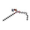  分子量: 758.060 Da / 分子数: 2 / 由来タイプ: 合成 / 式: C42H80NO8P / コメント: リン脂質*YM 分子量: 758.060 Da / 分子数: 2 / 由来タイプ: 合成 / 式: C42H80NO8P / コメント: リン脂質*YM#4: 糖 | ChemComp-MAN / |   タイプ: D-saccharide, alpha linking / 分子量: 180.156 Da / 分子数: 1 / 由来タイプ: 合成 / 式: C6H12O6 / タイプ: SUBJECT OF INVESTIGATION タイプ: D-saccharide, alpha linking / 分子量: 180.156 Da / 分子数: 1 / 由来タイプ: 合成 / 式: C6H12O6 / タイプ: SUBJECT OF INVESTIGATION研究の焦点であるリガンドがあるか | Y | Has protein modification | N | |
|---|
-実験情報
-実験
| 実験 | 手法: 電子顕微鏡法 |
|---|---|
| EM実験 | 試料の集合状態: PARTICLE / 3次元再構成法: 単粒子再構成法 |
- 試料調製
試料調製
| 構成要素 | 名称: Pmt4-Ccw5 complex / タイプ: COMPLEX / Entity ID: #1 / 由来: RECOMBINANT |
|---|---|
| 分子量 | 実験値: NO |
| 由来(天然) | 生物種: |
| 由来(組換発現) | 生物種: |
| 緩衝液 | pH: 7.5 |
| 試料 | 包埋: NO / シャドウイング: NO / 染色: NO / 凍結: YES |
| 急速凍結 | 凍結剤: ETHANE |
- 電子顕微鏡撮影
| 実験機器 |  モデル: Titan Krios / 画像提供: FEI Company |
|---|---|
| 顕微鏡 | モデル: TFS KRIOS |
| 電子銃 | 電子線源:  FIELD EMISSION GUN / 加速電圧: 300 kV / 照射モード: FLOOD BEAM FIELD EMISSION GUN / 加速電圧: 300 kV / 照射モード: FLOOD BEAM |
| 電子レンズ | モード: BRIGHT FIELD / 最大 デフォーカス(公称値): 1500 nm / 最小 デフォーカス(公称値): 800 nm |
| 撮影 | 電子線照射量: 50 e/Å2 フィルム・検出器のモデル: GATAN K3 BIOCONTINUUM (6k x 4k) |
- 解析
解析
| CTF補正 | タイプ: PHASE FLIPPING AND AMPLITUDE CORRECTION |
|---|---|
| 3次元再構成 | 解像度: 3.5 Å / 解像度の算出法: FSC 0.143 CUT-OFF / 粒子像の数: 48886 / 対称性のタイプ: POINT |
| 原子モデル構築 | プロトコル: AB INITIO MODEL |
