[English] 日本語
 Yorodumi
- EMDB-47566: Cryo-EM structure of Saccharomyces cerevisiae Pmt4-Ccw5 complex -
+ Open data
- Basic information
| Entry |  | |||||||||
|---|---|---|---|---|---|---|---|---|---|---|
| Title | Cryo-EM structure of Saccharomyces cerevisiae Pmt4-Ccw5 complex | |||||||||
 Map data Map data | structure of Saccharomyces cerevisiae Pmt4-Ccw5 complex | |||||||||
 Sample Sample |
| |||||||||
 Keywords Keywords | Pmt4 / O-mannosylation / MEMBRANE PROTEIN | |||||||||
| Function / homology |  Function and homology information Function and homology informationdolichyl-phosphate-mannose-protein mannosyltransferase Pmt4p homodimer complex / dolichyl-phosphate-mannose-protein mannosyltransferase / dolichyl-phosphate-mannose-protein mannosyltransferase activity / regulation of endoplasmic reticulum unfolded protein response / structural constituent of cell wall / fungal-type cell wall biogenesis / protein O-linked glycosylation via mannose / fungal-type cell wall organization / cellular bud tip / fungal-type cell wall ...dolichyl-phosphate-mannose-protein mannosyltransferase Pmt4p homodimer complex / dolichyl-phosphate-mannose-protein mannosyltransferase / dolichyl-phosphate-mannose-protein mannosyltransferase activity / regulation of endoplasmic reticulum unfolded protein response / structural constituent of cell wall / fungal-type cell wall biogenesis / protein O-linked glycosylation via mannose / fungal-type cell wall organization / cellular bud tip / fungal-type cell wall / fungal-type vacuole / protein O-linked glycosylation / cell periphery / endoplasmic reticulum membrane / endoplasmic reticulum / extracellular region / identical protein binding / plasma membrane Similarity search - Function | |||||||||
| Biological species |  | |||||||||
| Method | single particle reconstruction / cryo EM / Resolution: 3.2 Å | |||||||||
 Authors Authors | Du M / Yuan Z / Li H | |||||||||
| Funding support |  United States, 2 items United States, 2 items
| |||||||||
 Citation Citation |  Journal: Nat Commun / Year: 2025 Journal: Nat Commun / Year: 2025Title: Pmt4 recognizes two separate acceptor sites to O-mannosylate in the S/T-rich regions of substrate proteins. Authors: Minge Du / Zuanning Yuan / Amanda Kovach / Meinan Lyu / Huilin Li /  Abstract: Protein O-mannosyltransferases (PMTs) add mannose to serine/threonine (S/T)-rich proteins in the endoplasmic reticulum, facilitating proper folding and trafficking through the secretory pathway. ...Protein O-mannosyltransferases (PMTs) add mannose to serine/threonine (S/T)-rich proteins in the endoplasmic reticulum, facilitating proper folding and trafficking through the secretory pathway. These enzymes share a conserved architecture that includes a large transmembrane domain housing the catalytic pocket and a lumenal β-trefoil-folded MIR domain. Although S/T-rich regions in acceptor proteins are generally disordered, it remains unclear how PMTs selectively target these regions over other intrinsically disordered sequences. Here, using cryo-EM and X-ray crystallography, we demonstrate that the Saccharomyces cerevisiae Pmt4 dimer recognizes an S/T-rich peptide at two distinct sites. A groove above the catalytic pocket in the transmembrane domain binds the mannose-accepting S/T site, while the lumenal MIR domain engages an S/T-X-S/T motif. Notably, the substrate peptide is simultaneously bound by the catalytic pocket of one Pmt4 protomer and the MIR domain of the other, revealing an unexpected cooperative dual substrate recognition mechanism. This mechanism likely underpins the invariant dimeric architecture observed in all PMT family members. | |||||||||
| History |
|
- Structure visualization
| Supplemental images |
|---|
- Downloads & links
-EMDB archive
| Map data |  emd_47566.map.gz emd_47566.map.gz | 6.8 MB |  EMDB map data format EMDB map data format | |
|---|---|---|---|---|
| Header (meta data) |  emd-47566-v30.xml emd-47566-v30.xml emd-47566.xml emd-47566.xml | 18.9 KB 18.9 KB | Display Display |  EMDB header EMDB header |
| Images |  emd_47566.png emd_47566.png | 174.4 KB | ||
| Filedesc metadata |  emd-47566.cif.gz emd-47566.cif.gz | 6.4 KB | ||
| Others |  emd_47566_half_map_1.map.gz emd_47566_half_map_1.map.gz emd_47566_half_map_2.map.gz emd_47566_half_map_2.map.gz | 23.3 MB 23.3 MB | ||
| Archive directory |  http://ftp.pdbj.org/pub/emdb/structures/EMD-47566 http://ftp.pdbj.org/pub/emdb/structures/EMD-47566 ftp://ftp.pdbj.org/pub/emdb/structures/EMD-47566 ftp://ftp.pdbj.org/pub/emdb/structures/EMD-47566 | HTTPS FTP |
-Validation report
| Summary document |  emd_47566_validation.pdf.gz emd_47566_validation.pdf.gz | 777.3 KB | Display |  EMDB validaton report EMDB validaton report |
|---|---|---|---|---|
| Full document |  emd_47566_full_validation.pdf.gz emd_47566_full_validation.pdf.gz | 776.9 KB | Display | |
| Data in XML |  emd_47566_validation.xml.gz emd_47566_validation.xml.gz | 10.6 KB | Display | |
| Data in CIF |  emd_47566_validation.cif.gz emd_47566_validation.cif.gz | 12.4 KB | Display | |
| Arichive directory |  https://ftp.pdbj.org/pub/emdb/validation_reports/EMD-47566 https://ftp.pdbj.org/pub/emdb/validation_reports/EMD-47566 ftp://ftp.pdbj.org/pub/emdb/validation_reports/EMD-47566 ftp://ftp.pdbj.org/pub/emdb/validation_reports/EMD-47566 | HTTPS FTP |
-Related structure data
| Related structure data |  9e6iMC  9e61C  9e6vC  9e79C  9e7aC M: atomic model generated by this map C: citing same article ( |
|---|---|
| Similar structure data | Similarity search - Function & homology  F&H Search F&H Search |
- Links
Links
| EMDB pages |  EMDB (EBI/PDBe) / EMDB (EBI/PDBe) /  EMDataResource EMDataResource |
|---|
- Map
Map
| File |  Download / File: emd_47566.map.gz / Format: CCP4 / Size: 30.5 MB / Type: IMAGE STORED AS FLOATING POINT NUMBER (4 BYTES) Download / File: emd_47566.map.gz / Format: CCP4 / Size: 30.5 MB / Type: IMAGE STORED AS FLOATING POINT NUMBER (4 BYTES) | ||||||||||||||||||||||||||||||||||||
|---|---|---|---|---|---|---|---|---|---|---|---|---|---|---|---|---|---|---|---|---|---|---|---|---|---|---|---|---|---|---|---|---|---|---|---|---|---|
| Annotation | structure of Saccharomyces cerevisiae Pmt4-Ccw5 complex | ||||||||||||||||||||||||||||||||||||
| Projections & slices | Image control
Images are generated by Spider. | ||||||||||||||||||||||||||||||||||||
| Voxel size | X=Y=Z: 0.828 Å | ||||||||||||||||||||||||||||||||||||
| Density |
| ||||||||||||||||||||||||||||||||||||
| Symmetry | Space group: 1 | ||||||||||||||||||||||||||||||||||||
| Details | EMDB XML:
|
-Supplemental data
-Half map: Half Map B
| File | emd_47566_half_map_1.map | ||||||||||||
|---|---|---|---|---|---|---|---|---|---|---|---|---|---|
| Annotation | Half Map B | ||||||||||||
| Projections & Slices |
| ||||||||||||
| Density Histograms |
-Half map: Half Map A
| File | emd_47566_half_map_2.map | ||||||||||||
|---|---|---|---|---|---|---|---|---|---|---|---|---|---|
| Annotation | Half Map A | ||||||||||||
| Projections & Slices |
| ||||||||||||
| Density Histograms |
- Sample components
-Entire : Pmt4-Ccw5 complex
| Entire | Name: Pmt4-Ccw5 complex |
|---|---|
| Components |
|
-Supramolecule #1: Pmt4-Ccw5 complex
| Supramolecule | Name: Pmt4-Ccw5 complex / type: complex / ID: 1 / Parent: 0 / Macromolecule list: #1-#2 |
|---|---|
| Source (natural) | Organism:  |
-Macromolecule #1: Dolichyl-phosphate-mannose--protein mannosyltransferase 4
| Macromolecule | Name: Dolichyl-phosphate-mannose--protein mannosyltransferase 4 type: protein_or_peptide / ID: 1 / Number of copies: 2 / Enantiomer: LEVO EC number: dolichyl-phosphate-mannose-protein mannosyltransferase |
|---|---|
| Source (natural) | Organism:  |
| Molecular weight | Theoretical: 88.061945 KDa |
| Recombinant expression | Organism:  |
| Sequence | String: MSVPKKRNHG KLPPSTKDVD DPSLKYTKAA PKCEQVAEHW LLQPLPEPES RYSFWVTIVT LLAFAARFYK IWYPKEVVFD EVHFGKFAS YYLERSYFFD VHPPFAKMMI AFIGWLCGYD GSFKFDEIGY SYETHPAPYI AYRSFNAILG TLTVPIMFNT L KELNFRAI ...String: MSVPKKRNHG KLPPSTKDVD DPSLKYTKAA PKCEQVAEHW LLQPLPEPES RYSFWVTIVT LLAFAARFYK IWYPKEVVFD EVHFGKFAS YYLERSYFFD VHPPFAKMMI AFIGWLCGYD GSFKFDEIGY SYETHPAPYI AYRSFNAILG TLTVPIMFNT L KELNFRAI TCAFASLLVA IDTAHVTETR LILLDAILII SIAATMYCYV RFYKCQLRQP FTWSWYIWLH ATGLSLSFVI ST KYVGVMT YSAIGFAAVV NLWQLLDIKA GLSLRQFMRH FSKRLNGLVL IPFVIYLFWF WVHFTVLNTS GPGDAFMSAE FQE TLKDSP LSVDSKTVNY FDIITIKHQD TDAFLHSHLA RYPQRYEDGR ISSAGQQVTG YTHPDFNNQW EVLPPHGSDV GKGQ AVLLN QHIRLRHVAT DTYLLAHDVA SPFYPTNEEI TTVTLEEGDG ELYPETLFAF QPLKKSDEGH VLKSKTVSFR LFHVD TSVA LWTHNDELLP DWGFQQQEIN GNKKVIDPSN NWVVDEIVNL DEVRKVYIPK VVKPLPFLKK WIETQKSMFE HNNKLS SEH PFASEPYSWP GSLSGVSFWT NGDEKKQIYF IGNIIGWWFQ VISLAVFVGI IVADLITRHR GYYALNKMTR EKLYGPL MF FFVSWCCHYF PFFLMARQKF LHHYLPAHLI ACLFSGALWE VIFSDCKSLD LEKDEDISGA SYERNPKVYV KPYTVFLV C VSCAVAWFFV YFSPLVYGDV SLSPSEVVSR EWFDIELNFS K UniProtKB: Dolichyl-phosphate-mannose--protein mannosyltransferase 4 |
-Macromolecule #2: Cell wall mannoprotein CIS3
| Macromolecule | Name: Cell wall mannoprotein CIS3 / type: protein_or_peptide / ID: 2 / Number of copies: 1 / Enantiomer: LEVO |
|---|---|
| Source (natural) | Organism:  |
| Molecular weight | Theoretical: 1.556673 KDa |
| Recombinant expression | Organism:  |
| Sequence | String: TSTNATSSSC ATPSLK UniProtKB: Cell wall mannoprotein CIS3 |
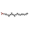
-Macromolecule #3: (3R)-3,31-dimethyl-7,11,15,19,23,27-hexamethylidenedotriacont-31-...
| Macromolecule | Name: (3R)-3,31-dimethyl-7,11,15,19,23,27-hexamethylidenedotriacont-31-en-1-yl dihydrogen phosphate type: ligand / ID: 3 / Number of copies: 1 / Formula: NNM |
|---|---|
| Molecular weight | Theoretical: 644.947 Da |
| Chemical component information |  ChemComp-NNM: |
-Experimental details
-Structure determination
| Method | cryo EM |
|---|---|
 Processing Processing | single particle reconstruction |
| Aggregation state | particle |
- Sample preparation
| Buffer | pH: 7.5 |
|---|---|
| Vitrification | Cryogen name: ETHANE |
- Electron microscopy
| Microscope | TFS KRIOS |
|---|---|
| Image recording | Film or detector model: GATAN K3 BIOCONTINUUM (6k x 4k) / Average electron dose: 50.0 e/Å2 |
| Electron beam | Acceleration voltage: 300 kV / Electron source:  FIELD EMISSION GUN FIELD EMISSION GUN |
| Electron optics | Illumination mode: FLOOD BEAM / Imaging mode: BRIGHT FIELD / Nominal defocus max: 1.5 µm / Nominal defocus min: 0.8 µm |
| Experimental equipment |  Model: Titan Krios / Image courtesy: FEI Company |
+ Image processing
-Atomic model buiding 1
| Refinement | Protocol: AB INITIO MODEL |
|---|---|
| Output model |  PDB-9e6i: |
 Movie
Movie Controller


 Z (Sec.)
 Y (Row.)
 X (Col.)


