ムービー
ムービー コントローラー


+ データを開く

- 基本情報
基本情報
| 登録情報 | データベース: PDB / ID: 9e6h | ||||||
|---|---|---|---|---|---|---|---|
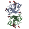
| タイトル | Cryo-EM structure of Maackia amurensis seed Leukoagglutinin (lectin), MASL | ||||||
 要素 要素 | Seed leukoagglutinin | ||||||
 キーワード キーワード | SUGAR BINDING PROTEIN / Seed Lectin / Maackia amurensis / N-linked glycosylation / Intersubunit Disulphide bridge / leukoagglutinin | ||||||
| 機能・相同性 |  機能・相同性情報 機能・相同性情報 | ||||||
| 生物種 |  Maackia amurensis (イヌエンジュ) Maackia amurensis (イヌエンジュ) | ||||||
| 手法 | 電子顕微鏡法 / 単粒子再構成法 / クライオ電子顕微鏡法 / 解像度: 2.84 Å | ||||||
 データ登録者 データ登録者 | Nayak, A.R. / Goldberg, G.S. / Temiakov, D. | ||||||
| 資金援助 |  米国, 1件 米国, 1件
| ||||||
 引用 引用 | ジャーナル: J Biol Chem / 年: 2025 タイトル: Maackia amurensis seed lectin structure and sequence comparison with other M. amurensis lectins. 著者: Ashok R Nayak / Cayla J Holdcraft / Ariel C Yin / Rachel E Nicoletto / Caifeng Zhao / Haiyan Zheng / Dmitry Temiakov / Gary S Goldberg / 要旨: Maackia amurensis lectins, including MASL, MAA, and MAL2, are widely utilized in biochemical and medicinal research. However, the structural and functional differences between these lectins have not ...Maackia amurensis lectins, including MASL, MAA, and MAL2, are widely utilized in biochemical and medicinal research. However, the structural and functional differences between these lectins have not been defined. Here, we present a high-resolution cryo-EM structure of MASL revealing that its tetrameric assembly is directed by two intersubunit disulfide bridges. These bridges, formed by C272 residues, are central to the dimer-of-dimers assembly of a MASL tetramer. This cryo-EM structure also identifies residues involved in stabilizing the dimer interface, multiple glycosylation sites, and calcium and manganese atoms in the sugar-binding pockets of MASL. Notably, our analysis reveals that Y250 in the carbohydrate-binding site of MASL adopts a flipped conformation, likely acting as a gatekeeper that obstructs access to noncognate substrates, a feature that may contribute to MASL's substrate specificity. Sequence analysis suggests that MAA is a truncated version of MASL, while MAL2 represents a homologous isoform. Unlike MASL, neither MAL2 nor MAA contains a cysteine residue required for disulfide bridge formation. Accordingly, analysis of these proteins using reducing and nonreducing SDS-PAGE confirms that the C272 residue in MASL drives intermolecular disulfide bridge formation. These findings provide critical insights into the unique structural features of MASL that distinguish it from other M. amurensis lectins, offering a foundation for further exploration of its biological and therapeutic potential. | ||||||
| 履歴 |
|
- 構造の表示
構造の表示
| 構造ビューア | 分子: MolmilJmol/JSmol |
|---|
- ダウンロードとリンク
-ダウンロード
| PDBx/mmCIF形式 | 9e6h.cif.gz | 209.3 KB | 表示 | PDBx/mmCIF形式 |
|---|---|---|---|---|
| PDB形式 | pdb9e6h.ent.gz | 166.7 KB | 表示 | PDB形式 |
| PDBx/mmJSON形式 | 9e6h.json.gz | ツリー表示 | PDBx/mmJSON形式 | |
| その他 |  その他のダウンロード その他のダウンロード |
-検証レポート
| 文書・要旨 | 9e6h_validation.pdf.gz | 1.9 MB | 表示 | wwPDB検証レポート |
|---|---|---|---|---|
| 文書・詳細版 | 9e6h_full_validation.pdf.gz | 1.9 MB | 表示 | |
| XML形式データ | 9e6h_validation.xml.gz | 41.8 KB | 表示 | |
| CIF形式データ | 9e6h_validation.cif.gz | 58.1 KB | 表示 | |
| アーカイブディレクトリ | https://data.pdbj.org/pub/pdb/validation_reports/e6/9e6hftp://data.pdbj.org/pub/pdb/validation_reports/e6/9e6h | HTTPS FTP |
-関連構造データ
-リンク
 PDBj
PDBj

- 集合体
集合体
| 登録構造単位 | 
|
|---|---|
| 1 |
|
-要素
-タンパク質 , 1種, 4分子 ABCD
| #1: タンパク質 | 分子量: 31321.480 Da / 分子数: 4 / 由来タイプ: 天然 / 由来: (天然) Maackia amurensis (イヌエンジュ) / 参照: UniProt: P0DKL3 |
|---|
-糖 , 3種, 16分子 
| #2: 多糖 | alpha-D-mannopyranose-(1-3)-beta-D-mannopyranose-(1-4)-2-acetamido-2-deoxy-beta-D-glucopyranose-(1- ...alpha-D-mannopyranose-(1-3)-beta-D-mannopyranose-(1-4)-2-acetamido-2-deoxy-beta-D-glucopyranose-(1-4)-2-acetamido-2-deoxy-beta-D-glucopyranose #3: 多糖 | alpha-D-mannopyranose-(1-3)-[alpha-D-mannopyranose-(1-6)]beta-D-mannopyranose-(1-4)-2-acetamido-2- ...alpha-D-mannopyranose-(1-3)-[alpha-D-mannopyranose-(1-6)]beta-D-mannopyranose-(1-4)-2-acetamido-2-deoxy-beta-D-glucopyranose-(1-4)-2-acetamido-2-deoxy-beta-D-glucopyranose #4: 糖 | ChemComp-NAG /  タイプ: D-saccharide, beta linking / 分子量: 221.208 Da / 分子数: 8 / 由来タイプ: 合成 / 式: C8H15NO6 / タイプ: SUBJECT OF INVESTIGATION タイプ: D-saccharide, beta linking / 分子量: 221.208 Da / 分子数: 8 / 由来タイプ: 合成 / 式: C8H15NO6 / タイプ: SUBJECT OF INVESTIGATION |
|---|
-非ポリマー , 2種, 8分子 

| #5: 化合物 | ChemComp-CA /  分子量: 40.078 Da / 分子数: 4 / 由来タイプ: 合成 / 式: Ca 分子量: 40.078 Da / 分子数: 4 / 由来タイプ: 合成 / 式: Ca#6: 化合物 | ChemComp-MN /  分子量: 54.938 Da / 分子数: 4 / 由来タイプ: 合成 / 式: Mn 分子量: 54.938 Da / 分子数: 4 / 由来タイプ: 合成 / 式: Mn |
|---|
-詳細
| 研究の焦点であるリガンドがあるか | Y |
|---|---|
| Has protein modification | Y |
-実験情報
-実験
| 実験 | 手法: 電子顕微鏡法 |
|---|---|
| EM実験 | 試料の集合状態: PARTICLE / 3次元再構成法: 単粒子再構成法 |
- 試料調製
試料調製
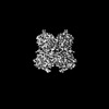
| 構成要素 | 名称: Cryo-EM map of Maackia amurensis seed Leukoagglutinin (lectin), MASL タイプ: COMPLEX / Entity ID: #1 / 由来: NATURAL |
|---|---|
| 分子量 | 値: 0.125 MDa / 実験値: YES |
| 由来(天然) | 生物種: Maackia amurensis (イヌエンジュ) |
| 由来(組換発現) | 生物種:  |
| 緩衝液 | pH: 7.9 詳細: 20 mM Tris-HCL (pH=7.9), 100 mM NaCL, 0.1 mM CaCl2, 0.1 mM MnCl2 |
| 試料 | 濃度: 0.4 mg/ml / 包埋: NO / シャドウイング: NO / 染色: NO / 凍結: YES / 詳細: 3 uM MASL monodisperse solution |
| 試料支持 | グリッドの材料: GOLD / グリッドのタイプ: UltrAuFoil R1.2/1.3 |
| 急速凍結 | 装置: FEI VITROBOT MARK IV / 凍結剤: ETHANE / 湿度: 95 % / 凍結前の試料温度: 277 K |
- 電子顕微鏡撮影
| 顕微鏡 | モデル: TFS GLACIOS / 詳細: Calibrated pixel size -0.93 |
|---|---|
| 電子銃 | 電子線源:  FIELD EMISSION GUN / 加速電圧: 200 kV / 照射モード: FLOOD BEAM FIELD EMISSION GUN / 加速電圧: 200 kV / 照射モード: FLOOD BEAM |
| 電子レンズ | モード: BRIGHT FIELD / 倍率(公称値): 150000 X / 最大 デフォーカス(公称値): 1200 nm / 最小 デフォーカス(公称値): 300 nm / Cs: 2.7 mm / C2レンズ絞り径: 50 µm / アライメント法: COMA FREE |
| 試料ホルダ | 凍結剤: NITROGEN 試料ホルダーモデル: FEI TITAN KRIOS AUTOGRID HOLDER |
| 撮影 | 電子線照射量: 60 e/Å2 フィルム・検出器のモデル: FEI FALCON IV (4k x 4k) 撮影したグリッド数: 1 / 実像数: 8543 詳細: Total number of frames - 40, Defocus range - -0.3 to -1.2 um in 0.1 increments, Output mode - TIFF, Autofocus recurrence - after centering, Drift measurement - once per grid square (Threshold - 0.4 nm/sec) |
| 電子光学装置 | 詳細: No Energy filter was used |
- 解析
解析
| EMソフトウェア |
| ||||||||||||||||||||||||||||
|---|---|---|---|---|---|---|---|---|---|---|---|---|---|---|---|---|---|---|---|---|---|---|---|---|---|---|---|---|---|
| CTF補正 | 詳細: CTFFIND 4.0 / タイプ: PHASE FLIPPING AND AMPLITUDE CORRECTION | ||||||||||||||||||||||||||||
| 粒子像の選択 | 選択した粒子像数: 8343216 | ||||||||||||||||||||||||||||
| 対称性 | 点対称性: C2 (2回回転対称) | ||||||||||||||||||||||||||||
| 3次元再構成 | 解像度: 2.84 Å / 解像度の算出法: FSC 0.143 CUT-OFF / 粒子像の数: 2656977 / クラス平均像の数: 2 / 対称性のタイプ: POINT | ||||||||||||||||||||||||||||
| 原子モデル構築 | プロトコル: FLEXIBLE FIT / 空間: REAL | ||||||||||||||||||||||||||||
| 原子モデル構築 | PDB-ID: 1DBN Accession code: 1DBN / Source name: PDB / タイプ: experimental model | ||||||||||||||||||||||||||||
| 拘束条件 |
|