Movie
Movie Controller

[English] 日本語
 Yorodumi
- EMDB-47565: Cryo-EM structure of Maackia amurensis seed Leukoagglutinin (lect... -
+ Open data

- Basic information
| Entry |  | |||||||||
|---|---|---|---|---|---|---|---|---|---|---|
| Title | Cryo-EM structure of Maackia amurensis seed Leukoagglutinin (lectin), MASL | |||||||||
 Map data Map data | ||||||||||
 Sample Sample |
| |||||||||
 Keywords Keywords | Seed Lectin / Maackia amurensis / N-linked glycosylation / Intersubunit Disulphide bridge / leukoagglutinin / SUGAR BINDING PROTEIN | |||||||||
| Function / homology |  Function and homology information Function and homology information | |||||||||
| Biological species |  Maackia amurensis (plant) Maackia amurensis (plant) | |||||||||
| Method | single particle reconstruction / cryo EM / Resolution: 2.84 Å | |||||||||
 Authors Authors | Nayak AR / Goldberg GS / Temiakov D / Sedmen J / Zamudio-Ochoa A | |||||||||
| Funding support |  United States, 1 items United States, 1 items
| |||||||||
 Citation Citation | Journal: J Biol Chem / Year: 2025 Title: Maackia amurensis seed lectin structure and sequence comparison with other M. amurensis lectins. Authors: Ashok R Nayak / Cayla J Holdcraft / Ariel C Yin / Rachel E Nicoletto / Caifeng Zhao / Haiyan Zheng / Dmitry Temiakov / Gary S Goldberg / Abstract: Maackia amurensis lectins, including MASL, MAA, and MAL2, are widely utilized in biochemical and medicinal research. However, the structural and functional differences between these lectins have not ...Maackia amurensis lectins, including MASL, MAA, and MAL2, are widely utilized in biochemical and medicinal research. However, the structural and functional differences between these lectins have not been defined. Here, we present a high-resolution cryo-EM structure of MASL revealing that its tetrameric assembly is directed by two intersubunit disulfide bridges. These bridges, formed by C272 residues, are central to the dimer-of-dimers assembly of a MASL tetramer. This cryo-EM structure also identifies residues involved in stabilizing the dimer interface, multiple glycosylation sites, and calcium and manganese atoms in the sugar-binding pockets of MASL. Notably, our analysis reveals that Y250 in the carbohydrate-binding site of MASL adopts a flipped conformation, likely acting as a gatekeeper that obstructs access to noncognate substrates, a feature that may contribute to MASL's substrate specificity. Sequence analysis suggests that MAA is a truncated version of MASL, while MAL2 represents a homologous isoform. Unlike MASL, neither MAL2 nor MAA contains a cysteine residue required for disulfide bridge formation. Accordingly, analysis of these proteins using reducing and nonreducing SDS-PAGE confirms that the C272 residue in MASL drives intermolecular disulfide bridge formation. These findings provide critical insights into the unique structural features of MASL that distinguish it from other M. amurensis lectins, offering a foundation for further exploration of its biological and therapeutic potential. | |||||||||
| History |
|
- Structure visualization
| Supplemental images |
|---|

- Downloads & links
-EMDB archive
| Map data | emd_47565.map.gz | 19.1 MB | EMDB map data format | |
|---|---|---|---|---|
| Header (meta data) | emd-47565-v30.xmlemd-47565.xml | 23 KB 23 KB | Display Display | EMDB header |
| FSC (resolution estimation) | emd_47565_fsc.xml | 7.1 KB | Display | FSC data file |
| Images |  emd_47565.png emd_47565.png | 82.1 KB | ||
| Filedesc metadata | emd-47565.cif.gz | 6.9 KB | ||
| Others | emd_47565_additional_1.map.gzemd_47565_half_map_1.map.gzemd_47565_half_map_2.map.gz | 19.9 MB 35.6 MB 35.6 MB | ||
| Archive directory |  http://ftp.pdbj.org/pub/emdb/structures/EMD-47565ftp://ftp.pdbj.org/pub/emdb/structures/EMD-47565 http://ftp.pdbj.org/pub/emdb/structures/EMD-47565ftp://ftp.pdbj.org/pub/emdb/structures/EMD-47565 | HTTPS FTP |
-Related structure data
| Related structure data | 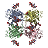 9e6hMC M: atomic model generated by this map C: citing same article ( |
|---|---|
| Similar structure data |
-Links
| EMDB pages | EMDB (EBI/PDBe) / EMDataResource |
|---|---|
| Related items in Molecule of the Month |


-Map
| File | Download / File: emd_47565.map.gz / Format: CCP4 / Size: 38.4 MB / Type: IMAGE STORED AS FLOATING POINT NUMBER (4 BYTES) | ||||||||||||||||||||||||||||||||||||
|---|---|---|---|---|---|---|---|---|---|---|---|---|---|---|---|---|---|---|---|---|---|---|---|---|---|---|---|---|---|---|---|---|---|---|---|---|---|
| Projections & slices | Image control
Images are generated by Spider. | ||||||||||||||||||||||||||||||||||||
| Voxel size | X=Y=Z: 0.93 Å | ||||||||||||||||||||||||||||||||||||
| Density |
| ||||||||||||||||||||||||||||||||||||
| Symmetry | Space group: 1 | ||||||||||||||||||||||||||||||||||||
| Details | EMDB XML:
|
 Z (Sec.)
 Y (Row.)
 X (Col.)


-Supplemental data
-Additional map: Sharpened map
| File | emd_47565_additional_1.map | ||||||||||||
|---|---|---|---|---|---|---|---|---|---|---|---|---|---|
| Annotation | Sharpened map | ||||||||||||
| Projections & Slices |
| ||||||||||||
| Density Histograms |


-Half map: #2
| File | emd_47565_half_map_1.map | ||||||||||||
|---|---|---|---|---|---|---|---|---|---|---|---|---|---|
| Projections & Slices |
| ||||||||||||
| Density Histograms |


-Half map: #1
| File | emd_47565_half_map_2.map | ||||||||||||
|---|---|---|---|---|---|---|---|---|---|---|---|---|---|
| Projections & Slices |
| ||||||||||||
| Density Histograms |


- Sample components
-Entire : Cryo-EM map of Maackia amurensis seed Leukoagglutinin (lectin), MASL
| Entire | Name: Cryo-EM map of Maackia amurensis seed Leukoagglutinin (lectin), MASL |
|---|---|
| Components |
|
-Supramolecule #1: Cryo-EM map of Maackia amurensis seed Leukoagglutinin (lectin), MASL
| Supramolecule | Name: Cryo-EM map of Maackia amurensis seed Leukoagglutinin (lectin), MASL type: complex / ID: 1 / Parent: 0 / Macromolecule list: #1 |
|---|---|
| Source (natural) | Organism: Maackia amurensis (plant) |
| Molecular weight | Theoretical: 125 KDa |
-Macromolecule #1: Seed leukoagglutinin
| Macromolecule | Name: Seed leukoagglutinin / type: protein_or_peptide / ID: 1 / Number of copies: 4 / Enantiomer: LEVO |
|---|---|
| Source (natural) | Organism: Maackia amurensis (plant) |
| Molecular weight | Theoretical: 31.32148 KDa |
| Sequence | String: MATSNSKPTQ VLLATFLTFF FLLLNNVNSS DELSFTINNF VPNEADLLFQ GEASVSSTGV LQLTRVENGQ PQQYSVGRAL YAAPVRIWD NTTGSVASFS TSFTFVVKAP NPDITSDGLA FYLAPPDSQI PSGSVSKYLG LFNNSNSDSS NQIVAVEFDT Y FGHSYDPW ...String: MATSNSKPTQ VLLATFLTFF FLLLNNVNSS DELSFTINNF VPNEADLLFQ GEASVSSTGV LQLTRVENGQ PQQYSVGRAL YAAPVRIWD NTTGSVASFS TSFTFVVKAP NPDITSDGLA FYLAPPDSQI PSGSVSKYLG LFNNSNSDSS NQIVAVEFDT Y FGHSYDPW DPNYRHIGID VNGIESIKTV QWDWINGGVA FATITYLAPN KTLIASLVYP SNQTTFSVAA SVDLKEILPE WV RVGFSAA TGYPTEVETH DVLSWSFTST LEANCDAATE NNVHIARYTA UniProtKB: Seed leukoagglutinin |
-Macromolecule #4: 2-acetamido-2-deoxy-beta-D-glucopyranose
| Macromolecule | Name: 2-acetamido-2-deoxy-beta-D-glucopyranose / type: ligand / ID: 4 / Number of copies: 8 / Formula: NAG |
|---|---|
| Molecular weight | Theoretical: 221.208 Da |
| Chemical component information |  ChemComp-NAG: |
-Macromolecule #5: CALCIUM ION
| Macromolecule | Name: CALCIUM ION / type: ligand / ID: 5 / Number of copies: 4 / Formula: CA |
|---|---|
| Molecular weight | Theoretical: 40.078 Da |
-Macromolecule #6: MANGANESE (II) ION
| Macromolecule | Name: MANGANESE (II) ION / type: ligand / ID: 6 / Number of copies: 4 / Formula: MN |
|---|---|
| Molecular weight | Theoretical: 54.938 Da |
-Experimental details
-Structure determination
| Method | cryo EM |
|---|---|
 Processing Processing | single particle reconstruction |
| Aggregation state | particle |
-Sample preparation
| Concentration | 0.4 mg/mL |
|---|---|
| Buffer | pH: 7.9 Details: 20 mM Tris-HCL (pH=7.9), 100 mM NaCL, 0.1 mM CaCl2, 0.1 mM MnCl2 |
| Grid | Model: UltrAuFoil R1.2/1.3 / Material: GOLD / Support film - Material: GOLD / Support film - topology: HOLEY / Pretreatment - Type: GLOW DISCHARGE |
| Vitrification | Cryogen name: ETHANE / Chamber humidity: 95 % / Chamber temperature: 277 K / Instrument: FEI VITROBOT MARK IV |
| Details | 3 uM MASL monodisperse solution |
- Electron microscopy
| Microscope | TFS GLACIOS |
|---|---|
| Specialist optics | Details: No Energy filter was used |
| Software | Name: EPU (ver. 3.6) |
| Details | Calibrated pixel size -0.93 |
| Image recording | Film or detector model: FEI FALCON IV (4k x 4k) / Number grids imaged: 1 / Number real images: 8543 / Average electron dose: 60.0 e/Å2 Details: Total number of frames - 40, Defocus range - -0.3 to -1.2 um in 0.1 increments, Output mode - TIFF, Autofocus recurrence - after centering, Drift measurement - once per grid square (Threshold - 0.4 nm/sec) |
| Electron beam | Acceleration voltage: 200 kV / Electron source:  FIELD EMISSION GUN FIELD EMISSION GUN |
| Electron optics | C2 aperture diameter: 50.0 µm / Illumination mode: FLOOD BEAM / Imaging mode: BRIGHT FIELD / Cs: 2.7 mm / Nominal defocus max: 1.2 µm / Nominal defocus min: 0.3 µm / Nominal magnification: 150000 |
| Sample stage | Specimen holder model: FEI TITAN KRIOS AUTOGRID HOLDER / Cooling holder cryogen: NITROGEN |