 Movie
Movie Controller

[English] 日本語
 Yorodumi
- PDB-9dzp: Photoactivation in Bacteriophytochromes, reference (dark) structu... -
+ Open data

- Basic information
| Entry | Database: PDB / ID: 9dzp | ||||||||||||
|---|---|---|---|---|---|---|---|---|---|---|---|---|---|
| Title | Photoactivation in Bacteriophytochromes, reference (dark) structure for the 100 ps time point | ||||||||||||
 Components Components | Photoreceptor-histidine kinase BphP | ||||||||||||
 Keywords Keywords | SIGNALING PROTEIN / Red-light receptor enzyme / Photosensory Core Domain | ||||||||||||
| Function / homology |  Function and homology information Function and homology informationosmosensory signaling via phosphorelay pathway / detection of visible light / phosphorelay response regulator activity / phosphorelay sensor kinase activity / histidine kinase / photoreceptor activity / protein kinase activator activity / regulation of DNA-templated transcription Similarity search - Function | ||||||||||||
| Biological species |  Stigmatella aurantiaca (bacteria) Stigmatella aurantiaca (bacteria) | ||||||||||||
| Method |  X-RAY DIFFRACTION / FREE ELECTRON LASER / FOURIER SYNTHESIS / Resolution: 1.93 Å X-RAY DIFFRACTION / FREE ELECTRON LASER / FOURIER SYNTHESIS / Resolution: 1.93 Å | ||||||||||||
 Authors Authors | Malla, T.N. / Stojkovic, E.A. / Schmidt, M. | ||||||||||||
| Funding support |  United States, 3items United States, 3items
| ||||||||||||
 Citation Citation | Journal: Commun Chem / Year: 2025 Title: Observation of early events in the photoactivation of Myxobacterial phytochrome using time-resolved serial femtosecond crystallography. Authors: Malla, T.N. / Aldama, L. / Leon, V. / Feliz, D. / Hu, H. / Thomas, I. / Cellini, A. / Wahlgren, W.Y. / Nimmrich, A. / Botha, S. / Sierra, R. / Hunter, M.S. / Poitevin, F. / Lisova, S. / ...Authors: Malla, T.N. / Aldama, L. / Leon, V. / Feliz, D. / Hu, H. / Thomas, I. / Cellini, A. / Wahlgren, W.Y. / Nimmrich, A. / Botha, S. / Sierra, R. / Hunter, M.S. / Poitevin, F. / Lisova, S. / Batyuk, A. / Gate, G. / Jernigan, R. / Kupitz, C.J. / Maj, P. / Meszaros, P. / Kurttila, M. / Monrroy, L. / Luo, F. / Owada, S. / Kang, J. / Slavov, C. / Maj, M. / Gautier, C. / Kashipathy, M. / Tolstikova, A. / Mariani, V. / Barty, A. / Moss, F. / Schwander, P. / Liu, H. / Boutet, S. / Fromme, P. / Takala, H. / Ihalainen, J.A. / Weierstall, U. / Westenhoff, S. / Stojkovic, E.A. / Schmidt, M. #1: Journal: Sci Adv / Year: 2024Title: Photoreception and signaling in bacterial phytochrome revealed by single-particle cryo-EM. Authors: Tek Narsingh Malla / Carolina Hernandez / Srinivasan Muniyappan / David Menendez / Dorina Bizhga / Joshua H Mendez / Peter Schwander / Emina A Stojković / Marius Schmidt / Abstract: Phytochromes are red-light photoreceptors discovered in plants with homologs in bacteria and fungi that regulate a variety of physiological responses. They display a reversible photocycle between two ...Phytochromes are red-light photoreceptors discovered in plants with homologs in bacteria and fungi that regulate a variety of physiological responses. They display a reversible photocycle between two distinct states: a red-light-absorbing Pr state and a far-red light-absorbing Pfr state. The photoconversion regulates the activity of an enzymatic domain, usually a histidine kinase (HK). The molecular mechanism that explains how light controls the HK activity is not understood because structures of unmodified bacterial phytochromes with HK activity are missing. Here, we report three cryo-electron microscopy structures of a wild-type bacterial phytochrome with HK activity determined as Pr and Pfr homodimers and as a Pr/Pfr heterodimer with individual subunits in distinct states. We propose that the Pr/Pfr heterodimer is a physiologically relevant signal transduction intermediate. Our results offer insight into the molecular mechanism that controls the enzymatic activity of the HK as part of a bacterial two-component system that perceives and transduces light signals. | ||||||||||||
| History |
|
- Structure visualization
| Structure viewer | Molecule: MolmilJmol/JSmol |
|---|
- Downloads & links
-Download
| PDBx/mmCIF format | 9dzp.cif.gz | 247.5 KB | Display | PDBx/mmCIF format |
|---|---|---|---|---|
| PDB format | pdb9dzp.ent.gz | 158.2 KB | Display | PDB format |
| PDBx/mmJSON format | 9dzp.json.gz | Tree view | PDBx/mmJSON format | |
| Others |  Other downloads Other downloads |
-Validation report
| Arichive directory | https://data.pdbj.org/pub/pdb/validation_reports/dz/9dzpftp://data.pdbj.org/pub/pdb/validation_reports/dz/9dzp | HTTPS FTP |
|---|
-Related structure data


-Links
 PDBj
PDBj


- Assembly
| Deposited unit | 
| ||||||||||||
|---|---|---|---|---|---|---|---|---|---|---|---|---|---|
| 1 |
| ||||||||||||
| Unit cell |
|
-Components
| #1: Protein | Mass: 51981.508 Da / Num. of mol.: 2 Source method: isolated from a genetically manipulated source Source: (gene. exp.) Stigmatella aurantiaca (bacteria) / Gene: STIAU_8420 / Production host: #2: Chemical | 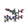  Mass: 584.662 Da / Num. of mol.: 2 / Source method: obtained synthetically / Formula: C33H36N4O6 / Feature type: SUBJECT OF INVESTIGATION Mass: 584.662 Da / Num. of mol.: 2 / Source method: obtained synthetically / Formula: C33H36N4O6 / Feature type: SUBJECT OF INVESTIGATION#3: Chemical | ChemComp-BEN / |   Mass: 120.152 Da / Num. of mol.: 1 / Source method: obtained synthetically / Formula: C7H8N2 Mass: 120.152 Da / Num. of mol.: 1 / Source method: obtained synthetically / Formula: C7H8N2#4: Water | ChemComp-HOH / |  Mass: 18.015 Da / Num. of mol.: 269 / Source method: isolated from a natural source / Formula: H2O Mass: 18.015 Da / Num. of mol.: 269 / Source method: isolated from a natural source / Formula: H2OHas ligand of interest | Y | Has protein modification | N | |
|---|
-Experimental details
-Experiment
| Experiment | Method: X-RAY DIFFRACTION / Number of used crystals: 1 |
|---|
- Sample preparation
| Crystal | Density Matthews: 2.76 Å3/Da / Density % sol: 55.47 % |
|---|---|
| Crystal grow | Temperature: 291 K / Method: batch mode Details: mother liquor: Hampton Crystal Screen Cryo HR2-122, #9 (PEG 4000), benzamidine HCl (3%) protein (60 mg/ml) to mother liquor 2:3. |
-Data collection
| Diffraction | Mean temperature: 293 K Ambient temp details: in vacuum, in hydrophobic grease matrix Serial crystal experiment: Y |
|---|---|
| Diffraction source | Source: FREE ELECTRON LASER / Site: SLAC LCLS / Beamline: CXI / Wavelength: 1.3 Å |
| Detector | Type: PSI JUNGFRAU 4M / Detector: PIXEL / Date: Feb 5, 2022 |
| Radiation | Monochromator: none / Protocol: SINGLE WAVELENGTH / Monochromatic (M) / Laue (L): M / Scattering type: x-ray |
| Radiation wavelength | Wavelength: 1.3 Å / Relative weight: 1 |
| Reflection | Resolution: 1.93→26.2 Å / Num. obs: 81351 / % possible obs: 100 % / Redundancy: 1644 % / Biso Wilson estimate: 39.02 Å2 / CC1/2: 0.99 / R split: 0.078 / Net I/σ(I): 7.04 |
| Reflection shell | Resolution: 1.93→2.01 Å / Redundancy: 1156 % / Mean I/σ(I) obs: 0.5 / Num. unique obs: 8910 / CC1/2: 0.12 / CC star: 0.46 / R split: 2.09 / % possible all: 100 |
| Serial crystallography measurement | Focal spot size: 5 µm2 / Pulse duration: 40 fsec. / Pulse photon energy: 9.3 keV / XFEL pulse repetition rate: 120 Hz |
| Serial crystallography sample delivery | Method: injection |
| Serial crystallography sample delivery injection | Carrier solvent: superlube / Crystal conc.: 0.1 / Description: viscous matrix injector / Flow rate: 3 µL/min / Injector diameter: 75 µm / Injector nozzle: toothpaste injector / Jet diameter: 75 µm / Power by: hydraulic / Preparation: folding at room temperature |
| Serial crystallography data reduction | Crystal hits: 239334 / Frames indexed: 108976 / XFEL run numbers: 21-43 |
- Processing
| Software |
| |||||||||||||||||||||||||||||||||||||||||||||||||||||||||||||||||||||||||||||||||||||||||||||||||||||||||||||||||||||||||||||||||||||||||||||||||||||||||||||||||||||||||||||||||||||||||||||
|---|---|---|---|---|---|---|---|---|---|---|---|---|---|---|---|---|---|---|---|---|---|---|---|---|---|---|---|---|---|---|---|---|---|---|---|---|---|---|---|---|---|---|---|---|---|---|---|---|---|---|---|---|---|---|---|---|---|---|---|---|---|---|---|---|---|---|---|---|---|---|---|---|---|---|---|---|---|---|---|---|---|---|---|---|---|---|---|---|---|---|---|---|---|---|---|---|---|---|---|---|---|---|---|---|---|---|---|---|---|---|---|---|---|---|---|---|---|---|---|---|---|---|---|---|---|---|---|---|---|---|---|---|---|---|---|---|---|---|---|---|---|---|---|---|---|---|---|---|---|---|---|---|---|---|---|---|---|---|---|---|---|---|---|---|---|---|---|---|---|---|---|---|---|---|---|---|---|---|---|---|---|---|---|---|---|---|---|---|---|---|
| Refinement | Method to determine structure: FOURIER SYNTHESIS / Resolution: 1.93→26.18 Å / SU ML: 0.2932 / Cross valid method: FREE R-VALUE / σ(F): 0.03 / Phase error: 28.6682 Stereochemistry target values: GeoStd + Monomer Library + CDL v1.2
| |||||||||||||||||||||||||||||||||||||||||||||||||||||||||||||||||||||||||||||||||||||||||||||||||||||||||||||||||||||||||||||||||||||||||||||||||||||||||||||||||||||||||||||||||||||||||||||
| Solvent computation | Shrinkage radii: 0.9 Å / VDW probe radii: 1.1 Å / Solvent model: FLAT BULK SOLVENT MODEL | |||||||||||||||||||||||||||||||||||||||||||||||||||||||||||||||||||||||||||||||||||||||||||||||||||||||||||||||||||||||||||||||||||||||||||||||||||||||||||||||||||||||||||||||||||||||||||||
| Displacement parameters | Biso mean: 48.5 Å2 | |||||||||||||||||||||||||||||||||||||||||||||||||||||||||||||||||||||||||||||||||||||||||||||||||||||||||||||||||||||||||||||||||||||||||||||||||||||||||||||||||||||||||||||||||||||||||||||
| Refinement step | Cycle: LAST / Resolution: 1.93→26.18 Å
| |||||||||||||||||||||||||||||||||||||||||||||||||||||||||||||||||||||||||||||||||||||||||||||||||||||||||||||||||||||||||||||||||||||||||||||||||||||||||||||||||||||||||||||||||||||||||||||
| Refine LS restraints |
| |||||||||||||||||||||||||||||||||||||||||||||||||||||||||||||||||||||||||||||||||||||||||||||||||||||||||||||||||||||||||||||||||||||||||||||||||||||||||||||||||||||||||||||||||||||||||||||
| LS refinement shell |
|
