 Movie
Movie Controller

[English] 日本語
 Yorodumi
- PDB-9dva: F-actin binding interface of alpha-E-catenin ABD (cadherin-cateni... -
+ Open data

- Basic information
| Entry | Database: PDB / ID: 9dva | |||||||||
|---|---|---|---|---|---|---|---|---|---|---|
| Title | F-actin binding interface of alpha-E-catenin ABD (cadherin-catenin complex) and afadin | |||||||||
 Components Components |
| |||||||||
 Keywords Keywords | STRUCTURAL PROTEIN / Cytoskeleton / cell adhesion | |||||||||
| Function / homology |  Function and homology information Function and homology informationpostsynaptic specialization organization / regulation of oligodendrocyte progenitor proliferation / radial glial cell differentiation / adherens junction maintenance / establishment of protein localization to plasma membrane / negative regulation of integrin-mediated signaling pathway / VEGFR2 mediated vascular permeability / positive regulation of cell-cell adhesion mediated by cadherin / protein localization to cell junction / Adherens junctions interactions ...postsynaptic specialization organization / regulation of oligodendrocyte progenitor proliferation / radial glial cell differentiation / adherens junction maintenance / establishment of protein localization to plasma membrane / negative regulation of integrin-mediated signaling pathway / VEGFR2 mediated vascular permeability / positive regulation of cell-cell adhesion mediated by cadherin / protein localization to cell junction / Adherens junctions interactions / RHO GTPases activate IQGAPs / epithelial cell-cell adhesion / Regulation of CDH1 Function / zonula adherens / establishment of endothelial intestinal barrier / Degradation of CDH1 / Regulation of CDH1 Function / gamma-catenin binding / positive regulation of cell-cell adhesion / dendrite arborization / gap junction assembly / neuroepithelial cell differentiation / LIM domain binding / Striated Muscle Contraction / cellular response to indole-3-methanol / pore complex assembly / positive regulation of dendrite extension / telencephalon development / vinculin binding / Myogenesis / bicellular tight junction assembly / cell-cell contact zone / flotillin complex / cell-cell adhesion mediated by cadherin / apical junction assembly / positive regulation of dendritic spine morphogenesis / catenin complex / negative regulation of cell motility / positive regulation of extrinsic apoptotic signaling pathway in absence of ligand / brain morphogenesis / positive regulation of smoothened signaling pathway / positive regulation of dendrite morphogenesis / positive regulation of mini excitatory postsynaptic potential / axon regeneration / tight junction / negative regulation of neuroblast proliferation / negative regulation of protein localization to nucleus / apical junction complex / pore complex / odontogenesis of dentin-containing tooth / establishment or maintenance of cell polarity / striated muscle thin filament / homeostasis of number of cells / skeletal muscle thin filament assembly / intercalated disc / regulation of postsynapse assembly / negative regulation of extrinsic apoptotic signaling pathway in absence of ligand / ovarian follicle development / skeletal muscle fiber development / stress fiber / somatodendritic compartment / presynaptic active zone membrane / cell adhesion molecule binding / excitatory synapse / acrosomal vesicle / negative regulation of cell migration / hippocampal mossy fiber to CA3 synapse / actin filament / adherens junction / cell-cell adhesion / cerebral cortex development / response to estrogen / beta-catenin binding / postsynaptic density membrane / male gonad development / Hydrolases; Acting on acid anhydrides; Acting on acid anhydrides to facilitate cellular and subcellular movement / small GTPase binding / apical part of cell / cell-cell junction / actin filament binding / cell junction / intracellular protein localization / regulation of cell population proliferation / regulation of protein localization / cell migration / lamellipodium / actin cytoskeleton / nuclear speck / cadherin binding / axon / hydrolase activity / positive regulation of gene expression / negative regulation of apoptotic process / structural molecule activity / glutamatergic synapse / Golgi apparatus / signal transduction / nucleoplasm / ATP binding / identical protein binding Similarity search - Function | |||||||||
| Biological species |   | |||||||||
| Method | ELECTRON MICROSCOPY / single particle reconstruction / cryo EM / Resolution: 3.1 Å | |||||||||
 Authors Authors | Gong, R. / Reynolds, M.J. / Alushin, G.M. | |||||||||
| Funding support |  United States, 2items United States, 2items
| |||||||||
 Citation Citation | Journal: bioRxiv / Year: 2024 Title: Afadin mediates cadherin-catenin complex clustering on F-actin linked to cooperative binding and filament curvature. Authors: Rui Gong / Matthew J Reynolds / Xiaoyu Sun / Gregory M Alushin / Abstract: The E-cadherin-β-catenin-αE-catenin (cadherin-catenin) complex couples the cytoskeletons of neighboring cells at adherens junctions (AJs) to mediate force transmission across epithelia. Mechanical ...The E-cadherin-β-catenin-αE-catenin (cadherin-catenin) complex couples the cytoskeletons of neighboring cells at adherens junctions (AJs) to mediate force transmission across epithelia. Mechanical force and auxiliary binding partners converge to stabilize the cadherin-catenin complex's inherently weak binding to actin filaments (F-actin) through unclear mechanisms. Here we show that afadin's coiled-coil (CC) domain and vinculin synergistically enhance the cadherin-catenin complex's F-actin engagement. The cryo-EM structure of an E-cadherin-β-catenin-αE-catenin-vinculin-afadin-CC supra-complex bound to F-actin reveals that afadin-CC bridges adjacent αE-catenin actin-binding domains along the filament, stabilizing flexible αE-catenin segments implicated in mechanical regulation. These cooperative binding contacts promote the formation of supra-complex clusters along F-actin. Additionally, cryo-EM variability analysis links supra-complex binding along individual F-actin strands to nanoscale filament curvature, a deformation mode associated with cytoskeletal forces. Collectively, this work elucidates a mechanistic framework by which vinculin and afadin tune cadherin-catenin complex-cytoskeleton coupling to support AJ function across varying mechanical regimes. | |||||||||
| History |
|
- Structure visualization
| Structure viewer | Molecule: MolmilJmol/JSmol |
|---|
- Downloads & links
-Download
| PDBx/mmCIF format | 9dva.cif.gz | 438.2 KB | Display | PDBx/mmCIF format |
|---|---|---|---|---|
| PDB format | pdb9dva.ent.gz | 344 KB | Display | PDB format |
| PDBx/mmJSON format | 9dva.json.gz | Tree view | PDBx/mmJSON format | |
| Others |  Other downloads Other downloads |
-Validation report
| Arichive directory | https://data.pdbj.org/pub/pdb/validation_reports/dv/9dvaftp://data.pdbj.org/pub/pdb/validation_reports/dv/9dva | HTTPS FTP |
|---|
-Related structure data
| Related structure data | 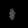 47194MC M: map data used to model this data C: citing same article ( |
|---|---|
| Similar structure data |


-Links
 PDBj
PDBj


- Assembly
| Deposited unit | 
|
|---|---|
| 1 |
|
-Components
| #1: Protein | Mass: 41875.633 Da / Num. of mol.: 5 / Source method: isolated from a natural source / Source: (natural) #2: Protein | Mass: 100110.078 Da / Num. of mol.: 2 Source method: isolated from a genetically manipulated source Source: (gene. exp.)  Homo sapiens (human) / References: UniProt: P26231 Homo sapiens (human) / References: UniProt: P26231#3: Protein | | Mass: 26150.164 Da / Num. of mol.: 1 Source method: isolated from a genetically manipulated source Source: (gene. exp.) Homo sapiens (human) / References: UniProt: Q9QZQ1#4: Chemical | ChemComp-ADP /   Mass: 427.201 Da / Num. of mol.: 5 / Source method: obtained synthetically / Formula: C10H15N5O10P2 / Feature type: SUBJECT OF INVESTIGATION / Comment: ADP, energy-carrying molecule*YM Mass: 427.201 Da / Num. of mol.: 5 / Source method: obtained synthetically / Formula: C10H15N5O10P2 / Feature type: SUBJECT OF INVESTIGATION / Comment: ADP, energy-carrying molecule*YM#5: Chemical | ChemComp-MG /   Mass: 24.305 Da / Num. of mol.: 5 / Source method: obtained synthetically / Formula: Mg / Feature type: SUBJECT OF INVESTIGATION Mass: 24.305 Da / Num. of mol.: 5 / Source method: obtained synthetically / Formula: Mg / Feature type: SUBJECT OF INVESTIGATIONHas ligand of interest | Y | Has protein modification | Y | |
|---|
-Experimental details
-Experiment
| Experiment | Method: ELECTRON MICROSCOPY |
|---|---|
| EM experiment | Aggregation state: FILAMENT / 3D reconstruction method: single particle reconstruction |
- Sample preparation
| Component | Name: F-actin binding interface of alpha-E-catenin ABD (cadherin-catenin complex) and afadin Type: COMPLEX / Entity ID: #1-#3 / Source: MULTIPLE SOURCES |
|---|---|
| Molecular weight | Value: 5.4 kDa/nm / Experimental value: YES |
| Source (natural) | Organism: Homo sapiens (human) |
| Source (recombinant) | Organism:  |
| Buffer solution | pH: 8 |
| Specimen | Embedding applied: NO / Shadowing applied: NO / Staining applied: NO / Vitrification applied: YES |
| Vitrification | Cryogen name: ETHANE |
- Electron microscopy imaging
| Experimental equipment |  Model: Titan Krios / Image courtesy: FEI Company |
|---|---|
| Microscopy | Model: FEI TITAN KRIOS |
| Electron gun | Electron source:  FIELD EMISSION GUN / Accelerating voltage: 300 kV / Illumination mode: FLOOD BEAM FIELD EMISSION GUN / Accelerating voltage: 300 kV / Illumination mode: FLOOD BEAM |
| Electron lens | Mode: BRIGHT FIELD / Nominal defocus max: 2800 nm / Nominal defocus min: 800 nm |
| Image recording | Electron dose: 61.26 e/Å2 / Film or detector model: GATAN K2 SUMMIT (4k x 4k) |
- Processing
| CTF correction | Type: PHASE FLIPPING AND AMPLITUDE CORRECTION |
|---|---|
| 3D reconstruction | Resolution: 3.1 Å / Resolution method: FSC 0.143 CUT-OFF / Num. of particles: 99745 / Symmetry type: POINT |
