National Institutes of Health/National Institute of General Medical Sciences (NIH/NIGMS)
R01GM141044
United States
Other private
G-2020-14047
Citation
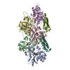
Journal: bioRxiv / Year: 2024 Title: Afadin mediates cadherin-catenin complex clustering on F-actin linked to cooperative binding and filament curvature. Authors: Rui Gong / Matthew J Reynolds / Xiaoyu Sun / Gregory M Alushin / Abstract: The E-cadherin-β-catenin-αE-catenin (cadherin-catenin) complex couples the cytoskeletons of neighboring cells at adherens junctions (AJs) to mediate force transmission across epithelia. Mechanical ...The E-cadherin-β-catenin-αE-catenin (cadherin-catenin) complex couples the cytoskeletons of neighboring cells at adherens junctions (AJs) to mediate force transmission across epithelia. Mechanical force and auxiliary binding partners converge to stabilize the cadherin-catenin complex's inherently weak binding to actin filaments (F-actin) through unclear mechanisms. Here we show that afadin's coiled-coil (CC) domain and vinculin synergistically enhance the cadherin-catenin complex's F-actin engagement. The cryo-EM structure of an E-cadherin-β-catenin-αE-catenin-vinculin-afadin-CC supra-complex bound to F-actin reveals that afadin-CC bridges adjacent αE-catenin actin-binding domains along the filament, stabilizing flexible αE-catenin segments implicated in mechanical regulation. These cooperative binding contacts promote the formation of supra-complex clusters along F-actin. Additionally, cryo-EM variability analysis links supra-complex binding along individual F-actin strands to nanoscale filament curvature, a deformation mode associated with cytoskeletal forces. Collectively, this work elucidates a mechanistic framework by which vinculin and afadin tune cadherin-catenin complex-cytoskeleton coupling to support AJ function across varying mechanical regimes.
In the structure databanks used in Yorodumi, some data are registered as the other names, "COVID-19 virus" and "2019-nCoV". Here are the details of the virus and the list of structure data.
Jan 31, 2019. EMDB accession codes are about to change! (news from PDBe EMDB page)
EMDB accession codes are about to change! (news from PDBe EMDB page)
The allocation of 4 digits for EMDB accession codes will soon come to an end. Whilst these codes will remain in use, new EMDB accession codes will include an additional digit and will expand incrementally as the available range of codes is exhausted. The current 4-digit format prefixed with “EMD-” (i.e. EMD-XXXX) will advance to a 5-digit format (i.e. EMD-XXXXX), and so on. It is currently estimated that the 4-digit codes will be depleted around Spring 2019, at which point the 5-digit format will come into force.
The EM Navigator/Yorodumi systems omit the EMD- prefix.
Related info.:Q: What is EMD? / ID/Accession-code notation in Yorodumi/EM Navigator
Yorodumi is a browser for structure data from EMDB, PDB, SASBDB, etc.
This page is also the successor to EM Navigator detail page, and also detail information page/front-end page for Omokage search.
The word "yorodu" (or yorozu) is an old Japanese word meaning "ten thousand". "mi" (miru) is to see.
Related info.:EMDB / PDB / SASBDB / Comparison of 3 databanks / Yorodumi Search / Aug 31, 2016. New EM Navigator & Yorodumi / Yorodumi Papers / Jmol/JSmol / Function and homology information / Changes in new EM Navigator and Yorodumi
 Movie
Movie Controller

 Yorodumi
 Open data
 Basic information
 Map data
 Sample
 Keywords
 Function and homology information
 Homo sapiens (human) /

 Authors
 United States, 2 items
  Citation
 Structure visualization
 Downloads & links
 emd_47194.png
 http://ftp.pdbj.org/pub/emdb/structures/EMD-47194
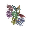


 Z (Sec.)
 Y (Row.)
 X (Col.)


 Sample components
 Processing
 Electron microscopy
 FIELD EMISSION GUN