ムービー
ムービー コントローラー


+ データを開く

- 基本情報
基本情報
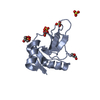
| 登録情報 | データベース: PDB / ID: 9dkz | ||||||||||||||||||
|---|---|---|---|---|---|---|---|---|---|---|---|---|---|---|---|---|---|---|---|
| タイトル | In situ microED structure of the Eosinophil major basic protein-1 | ||||||||||||||||||
 要素 要素 | Bone marrow proteoglycan | ||||||||||||||||||
 キーワード キーワード | IMMUNE SYSTEM / Effector / Nanocrystal / In-situ / Intracellular | ||||||||||||||||||
| 機能・相同性 |  機能・相同性情報 機能・相同性情報extracellular matrix structural constituent conferring compression resistance / defense response to nematode / negative regulation of macrophage cytokine production / negative regulation of interleukin-10 production / positive regulation of interleukin-4 production / transport vesicle / heparin binding / carbohydrate binding / extracellular matrix / ficolin-1-rich granule lumen ...extracellular matrix structural constituent conferring compression resistance / defense response to nematode / negative regulation of macrophage cytokine production / negative regulation of interleukin-10 production / positive regulation of interleukin-4 production / transport vesicle / heparin binding / carbohydrate binding / extracellular matrix / ficolin-1-rich granule lumen / defense response to bacterium / immune response / Neutrophil degranulation / extracellular exosome / extracellular region 類似検索 - 分子機能 | ||||||||||||||||||
| 生物種 |  Homo sapiens (ヒト) Homo sapiens (ヒト) | ||||||||||||||||||
| 手法 | 電子線結晶学 / クライオ電子顕微鏡法 / 解像度: 3.2 Å | ||||||||||||||||||
 データ登録者 データ登録者 | Yang, J.E. / Bingman, C.A. / Mitchell, J. / Mosher, D. / Wright, E.R. | ||||||||||||||||||
| 資金援助 |  米国, 5件 米国, 5件
| ||||||||||||||||||
 引用 引用 | ジャーナル: bioRxiv / 年: 2024 タイトル: In situ crystalline structure of the human eosinophil major basic protein-1. 著者: Jie E Yang / Joshua M Mitchell / Craig A Bingman / Deane F Mosher / Elizabeth R Wright / 要旨: Eosinophils are white blood cells that participate in innate immune responses and have an essential role in the pathogenesis of inflammatory and neoplastic disorders. Upon activation, eosinophils ...Eosinophils are white blood cells that participate in innate immune responses and have an essential role in the pathogenesis of inflammatory and neoplastic disorders. Upon activation, eosinophils release cytotoxic proteins such as major basic protein-1 (MBP-1) from cytoplasmic secretory granules (SGr) wherein MBP-1 is stored as nanocrystals. How the MBP-1 nanocrystalline core is formed, stabilized, and subsequently mobilized remains unknown. Here, we report the structure of crystalline MBP-1 within SGrs of human eosinophils. The structure reveals a mechanism for intragranular crystal packing and stabilization of MBP-1 via a structurally conserved loop region that is associated with calcium-dependent carbohydrate binding in other C-type lectin (CTL) proteins. Single-cell and single-SGr profiling correlating real-space three-dimensional information from cellular montage cryo-electron tomography (cryo-ET) and microcrystal electron diffraction (MicroED) data obtained from non-activated and IL33-activated eosinophils revealed activation-dependent crystal expansion and extrusion of expanded crystals from SGr. These results suggest that MBP-1 crystals play a dynamic role in the release of SGr contents. Collectively, this research demonstrates the importance of macromolecular structure determination. | ||||||||||||||||||
| 履歴 |
|
- 構造の表示
構造の表示
| 構造ビューア | 分子: MolmilJmol/JSmol |
|---|
- ダウンロードとリンク
-ダウンロード
| PDBx/mmCIF形式 | 9dkz.cif.gz | 61 KB | 表示 | PDBx/mmCIF形式 |
|---|---|---|---|---|
| PDB形式 | pdb9dkz.ent.gz | 41.2 KB | 表示 | PDB形式 |
| PDBx/mmJSON形式 | 9dkz.json.gz | ツリー表示 | PDBx/mmJSON形式 | |
| その他 |  その他のダウンロード その他のダウンロード |
-検証レポート
| アーカイブディレクトリ | https://data.pdbj.org/pub/pdb/validation_reports/dk/9dkzftp://data.pdbj.org/pub/pdb/validation_reports/dk/9dkz | HTTPS FTP |
|---|
-関連構造データ
| 関連構造データ | |
|---|---|
| 類似構造データ |

-リンク
 PDBj
PDBj


- 集合体
集合体
| 登録構造単位 | 
| ||||||||||||
|---|---|---|---|---|---|---|---|---|---|---|---|---|---|
| 1 |
| ||||||||||||
| 単位格子 |
|
-要素
| #1: タンパク質 | 分子量: 13694.769 Da / 分子数: 1 / 由来タイプ: 組換発現 詳細: The major basic protein-1 structure was directly determined from intragranular nanocrystals inside unperturbed human eosinophil cells obtained from donors. 由来: (組換発現) Homo sapiens (ヒト) / 組織: blood / Cell: eosinophil / 遺伝子: PRG2, MBP / 発現宿主: Homo sapiens (ヒト) / 参照: UniProt: P13727 |
|---|---|
| #2: 水 | ChemComp-HOH /  分子量: 18.015 Da / 分子数: 1 / 由来タイプ: 天然 / 式: H2O 分子量: 18.015 Da / 分子数: 1 / 由来タイプ: 天然 / 式: H2O |
| Has protein modification | Y |
-実験情報
-実験
| 実験 | 手法: 電子線結晶学 |
|---|---|
| EM実験 | 試料の集合状態: CELL / 3次元再構成法: 電子線結晶学 |
- 試料調製
試料調製
| 構成要素 | 名称: In situ microED structure of the Eosinophil major basic protein-1 タイプ: CELL 詳細: Mature human eosinophil cells were collected and purified from human donor blood. This was followed by eosinophil cell deposition on EM grids, grid plunge-freezing, and cryo-FIB milling of ...詳細: Mature human eosinophil cells were collected and purified from human donor blood. This was followed by eosinophil cell deposition on EM grids, grid plunge-freezing, and cryo-FIB milling of individual eosinophil cells. Cryo-FIB milling of the cells exposed unperturbed cytosolic secretory granules, inside which nanocrystals of the major basic protein-1 were located. Micro-ED data was collected on these crystals for in-situ structure determination. Entity ID: #1 / 由来: NATURAL |
|---|---|
| 由来(天然) | 生物種: Homo sapiens (ヒト) / 細胞内の位置: cytoplasm / Organelle: mature eosinophil secretory granule / 組織: blood |
| 緩衝液 | pH: 7 / 詳細: secretory granule matrix |
| 試料 | 濃度: 1000000 mg/ml / 包埋: NO / シャドウイング: NO / 染色: NO / 凍結: YES 詳細: Human eosinophil cells were collected from human donor blood. The cells were rested in 1640 RPMI medium supplemented with 0.1% human serum albumin, before direct deposition onto gold EM grids ...詳細: Human eosinophil cells were collected from human donor blood. The cells were rested in 1640 RPMI medium supplemented with 0.1% human serum albumin, before direct deposition onto gold EM grids and plunge freezing. Subsequently, cryo-FIB milling was used to expose unperturbed intragranular major basic protien-1 nanocrystals present within the cytosolic secretory granules. |
| 試料支持 | 詳細: 10 mA / グリッドの材料: GOLD / グリッドのサイズ: 200 divisions/in. / グリッドのタイプ: Quantifoil |
| 急速凍結 | 装置: LEICA EM GP / 凍結剤: ETHANE / 湿度: 95 % / 凍結前の試料温度: 310.15 K / 詳細: Blot time of 6~8 sec, single-side back blotting |
-データ収集
| 実験機器 |  モデル: Titan Krios / 画像提供: FEI Company | ||||||||||||||||||||||||||||||||||||||||||||||||||||||||||||||||||||||||||||||||||||||||||||||||||||||||||||||||||||||||||||||||||||||||||||||||||||||||||||||||||||||||||||||||||||
|---|---|---|---|---|---|---|---|---|---|---|---|---|---|---|---|---|---|---|---|---|---|---|---|---|---|---|---|---|---|---|---|---|---|---|---|---|---|---|---|---|---|---|---|---|---|---|---|---|---|---|---|---|---|---|---|---|---|---|---|---|---|---|---|---|---|---|---|---|---|---|---|---|---|---|---|---|---|---|---|---|---|---|---|---|---|---|---|---|---|---|---|---|---|---|---|---|---|---|---|---|---|---|---|---|---|---|---|---|---|---|---|---|---|---|---|---|---|---|---|---|---|---|---|---|---|---|---|---|---|---|---|---|---|---|---|---|---|---|---|---|---|---|---|---|---|---|---|---|---|---|---|---|---|---|---|---|---|---|---|---|---|---|---|---|---|---|---|---|---|---|---|---|---|---|---|---|---|---|---|---|---|
| 顕微鏡 | モデル: TFS KRIOS | ||||||||||||||||||||||||||||||||||||||||||||||||||||||||||||||||||||||||||||||||||||||||||||||||||||||||||||||||||||||||||||||||||||||||||||||||||||||||||||||||||||||||||||||||||||
| 電子銃 | 電子線源:  FIELD EMISSION GUN / 加速電圧: 300 kV / 照射モード: FLOOD BEAM FIELD EMISSION GUN / 加速電圧: 300 kV / 照射モード: FLOOD BEAM | ||||||||||||||||||||||||||||||||||||||||||||||||||||||||||||||||||||||||||||||||||||||||||||||||||||||||||||||||||||||||||||||||||||||||||||||||||||||||||||||||||||||||||||||||||||
| 電子レンズ | モード: DIFFRACTION / 最大 デフォーカス(公称値): 0 nm / 最小 デフォーカス(公称値): 0 nm / Calibrated defocus min: 0 nm / 最大 デフォーカス(補正後): 0 nm / Cs: 2.7 mm / C2レンズ絞り径: 50 µm / アライメント法: COMA FREE | ||||||||||||||||||||||||||||||||||||||||||||||||||||||||||||||||||||||||||||||||||||||||||||||||||||||||||||||||||||||||||||||||||||||||||||||||||||||||||||||||||||||||||||||||||||
| 試料ホルダ | 凍結剤: NITROGEN 試料ホルダーモデル: FEI TITAN KRIOS AUTOGRID HOLDER 最高温度: 100 K / 最低温度: 85 K | ||||||||||||||||||||||||||||||||||||||||||||||||||||||||||||||||||||||||||||||||||||||||||||||||||||||||||||||||||||||||||||||||||||||||||||||||||||||||||||||||||||||||||||||||||||
| 撮影 | 平均露光時間: 1 sec. / 電子線照射量: 0.15 e/Å2 / フィルム・検出器のモデル: FEI CETA (4k x 4k) / Num. of diffraction images: 40 / 撮影したグリッド数: 3 詳細: The final dataset was merged from six crystals, each with 40 images. Tilt was -20 to +20 degrees, 1 degree per frame, 1 second per frame | ||||||||||||||||||||||||||||||||||||||||||||||||||||||||||||||||||||||||||||||||||||||||||||||||||||||||||||||||||||||||||||||||||||||||||||||||||||||||||||||||||||||||||||||||||||
| EM回折 シェル | 解像度: 3.2→28.93 Å / フーリエ空間範囲: 95.6 % / 多重度: 7.5 / 構造因子数: 1912 / 位相残差: 26.33 ° | ||||||||||||||||||||||||||||||||||||||||||||||||||||||||||||||||||||||||||||||||||||||||||||||||||||||||||||||||||||||||||||||||||||||||||||||||||||||||||||||||||||||||||||||||||||
| EM回折 統計 | フーリエ空間範囲: 95.6 % / 再高解像度: 3.2 Å / 測定した強度の数: 24722 / 構造因子数: 3303 / 位相誤差: 26.33 ° / 位相誤差の除外基準: 0 / Rmerge: 67.29 | ||||||||||||||||||||||||||||||||||||||||||||||||||||||||||||||||||||||||||||||||||||||||||||||||||||||||||||||||||||||||||||||||||||||||||||||||||||||||||||||||||||||||||||||||||||
| 反射 | 最高解像度: 3.2 Å / Num. obs: 1912 / % possible obs: 96.1 % / Biso Wilson estimate: 85.01 Å2 / CC1/2: 0.931 / Rmerge(I) obs: 0.69 / Rrim(I) all: 0.719 / Net I/σ(I): 3.2 / Num. measured all: 24817 | ||||||||||||||||||||||||||||||||||||||||||||||||||||||||||||||||||||||||||||||||||||||||||||||||||||||||||||||||||||||||||||||||||||||||||||||||||||||||||||||||||||||||||||||||||||
| 反射 シェル | Diffraction-ID: 1
|
- 解析
解析
| ソフトウェア |
| ||||||||||||||||||||||||||||||||||||||||||
|---|---|---|---|---|---|---|---|---|---|---|---|---|---|---|---|---|---|---|---|---|---|---|---|---|---|---|---|---|---|---|---|---|---|---|---|---|---|---|---|---|---|---|---|
| EMソフトウェア |
| ||||||||||||||||||||||||||||||||||||||||||
| EM 3D crystal entity | ∠α: 90 ° / ∠β: 90 ° / ∠γ: 90 ° / A: 31.24 Å / B: 57.87 Å / C: 59.06 Å / 空間群名: P22121 / 空間群番号: 18 | ||||||||||||||||||||||||||||||||||||||||||
| CTF補正 | タイプ: NONE | ||||||||||||||||||||||||||||||||||||||||||
| 3次元再構成 | 解像度: 3.2 Å / 解像度の算出法: DIFFRACTION PATTERN/LAYERLINES / 対称性のタイプ: 3D CRYSTAL | ||||||||||||||||||||||||||||||||||||||||||
| 原子モデル構築 | プロトコル: OTHER / 空間: RECIPROCAL 詳細: Alternating rounds of phenix.refine and map filling in Coot | ||||||||||||||||||||||||||||||||||||||||||
| 原子モデル構築 | 3D fitting-ID: 1 / Chain-ID: A / Chain residue range: 106-222
| ||||||||||||||||||||||||||||||||||||||||||
| 精密化 | 解像度: 3.2→28.93 Å / SU ML: 0.2682 / 交差検証法: FREE R-VALUE / 位相誤差: 26.171 立体化学のターゲット値: GeoStd + Monomer Library + CDL v1.2
| ||||||||||||||||||||||||||||||||||||||||||
| 溶媒の処理 | 減衰半径: 0.9 Å / VDWプローブ半径: 1.1 Å / 溶媒モデル: FLAT BULK SOLVENT MODEL | ||||||||||||||||||||||||||||||||||||||||||
| 原子変位パラメータ | Biso mean: 91.79 Å2 | ||||||||||||||||||||||||||||||||||||||||||
| 拘束条件 |
| ||||||||||||||||||||||||||||||||||||||||||
| LS精密化 シェル | Refine-ID: ELECTRON CRYSTALLOGRAPHY
|