Movie
Movie Controller


+ Open data

- Basic information
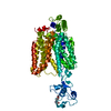
| Entry | Database: PDB / ID: 9dkc | ||||||
|---|---|---|---|---|---|---|---|
| Title | Structure of URAT1 in complex with TD-3 | ||||||
 Components Components | URAT1 | ||||||
 Keywords Keywords | TRANSPORT PROTEIN / Membrane protein / membrane transporter | ||||||
| Function / homology | :  Function and homology information Function and homology information | ||||||
| Biological species |  Homo sapiens (human) Homo sapiens (human) | ||||||
| Method | ELECTRON MICROSCOPY / single particle reconstruction / cryo EM / Resolution: 2.55 Å | ||||||
 Authors Authors | Suo, Y. / Fedor, J.G. / Lee, S.-Y. | ||||||
| Funding support |  United States, 1items United States, 1items
| ||||||
 Citation Citation | Journal: Nat Commun / Year: 2025 Title: Molecular basis of the urate transporter URAT1 inhibition by gout drugs. Authors: Yang Suo / Justin G Fedor / Han Zhang / Kalina Tsolova / Xiaoyu Shi / Kedar Sharma / Shweta Kumari / Mario Borgnia / Peng Zhan / Wonpil Im / Seok-Yong Lee /  Abstract: Hyperuricemia is a condition when uric acid, a waste product of purine metabolism, accumulates in the blood. Untreated hyperuricemia can lead to crystal formation of monosodium urate in the joints, ...Hyperuricemia is a condition when uric acid, a waste product of purine metabolism, accumulates in the blood. Untreated hyperuricemia can lead to crystal formation of monosodium urate in the joints, causing a painful inflammatory disease known as gout. These conditions are associated with many other diseases and affect a significant and increasing proportion of the population. The human urate transporter 1 (URAT1) is responsible for the reabsorption of ~90% of uric acid in the kidneys back into the blood, making it a primary target for treating hyperuricemia and gout. Despite decades of research and development, clinically available URAT1 inhibitors have limitations because the molecular basis of URAT1 inhibition by gout drugs remains unknown. Here we present cryo-electron microscopy structures of URAT1 alone and in complex with three clinically relevant inhibitors: benzbromarone, lesinurad, and the recently developed compound TD-3. Together with functional experiments and molecular dynamics simulations, we reveal that these inhibitors bind selectively to URAT1 in inward-open states. Furthermore, we discover differences in the inhibitor-dependent URAT1 conformations as well as interaction networks, which contribute to drug specificity. Our findings illuminate a general theme for URAT1 inhibition, paving the way for the design of next-generation URAT1 inhibitors in the treatment of gout and hyperuricemia. | ||||||
| History |
|
- Structure visualization
| Structure viewer | Molecule: MolmilJmol/JSmol |
|---|
- Downloads & links
-Download
| PDBx/mmCIF format | 9dkc.cif.gz | 193.7 KB | Display | PDBx/mmCIF format |
|---|---|---|---|---|
| PDB format | pdb9dkc.ent.gz | Display | PDB format | |
| PDBx/mmJSON format | 9dkc.json.gz | Tree view | PDBx/mmJSON format | |
| Others |  Other downloads Other downloads |
-Validation report
| Arichive directory | https://data.pdbj.org/pub/pdb/validation_reports/dk/9dkcftp://data.pdbj.org/pub/pdb/validation_reports/dk/9dkc | HTTPS FTP |
|---|
-Related structure data
| Related structure data |  46951MC  9dk9C  9dkaC  9dkbC M: map data used to model this data C: citing same article ( |
|---|---|
| Similar structure data |


-Links
 PDBj
PDBj- Assembly
| Deposited unit | 
|
|---|---|
| 1 |
|
-Components
| #1: Protein | Mass: 55379.844 Da / Num. of mol.: 1 Source method: isolated from a genetically manipulated source Source: (gene. exp.) Homo sapiens (human) / Production host: Homo sapiens (human) |
|---|---|
| #2: Chemical | ChemComp-A1A45 / Mass: 456.356 Da / Num. of mol.: 1 / Source method: obtained synthetically / Formula: C21H18BrN3O2S |
| Has ligand of interest | Y |
| Has protein modification | Y |
-Experimental details
-Experiment
| Experiment | Method: ELECTRON MICROSCOPY |
|---|---|
| EM experiment | Aggregation state: PARTICLE / 3D reconstruction method: single particle reconstruction |
- Sample preparation
| Component | Name: URAT1 / Type: COMPLEX / Entity ID: #1 / Source: RECOMBINANT | ||||||||||||||||||||
|---|---|---|---|---|---|---|---|---|---|---|---|---|---|---|---|---|---|---|---|---|---|
| Molecular weight | Value: 0.055 MDa / Experimental value: NO | ||||||||||||||||||||
| Source (natural) | Organism: Homo sapiens (human) | ||||||||||||||||||||
| Source (recombinant) | Organism: Homo sapiens (human) | ||||||||||||||||||||
| Buffer solution | pH: 8 | ||||||||||||||||||||
| Buffer component |
| ||||||||||||||||||||
| Specimen | Conc.: 10 mg/ml / Embedding applied: NO / Shadowing applied: NO / Staining applied: NO / Vitrification applied: YES / Details: Monodisperse sample | ||||||||||||||||||||
| Specimen support | Grid type: Quantifoil R1.2/1.3 | ||||||||||||||||||||
| Vitrification | Instrument: LEICA EM GP / Cryogen name: ETHANE / Humidity: 95 % / Chamber temperature: 280 K |
- Electron microscopy imaging
| Experimental equipment |  Model: Titan Krios / Image courtesy: FEI Company |
|---|---|
| Microscopy | Model: TFS KRIOS |
| Electron gun | Electron source:  FIELD EMISSION GUN / Accelerating voltage: 300 kV / Illumination mode: FLOOD BEAM FIELD EMISSION GUN / Accelerating voltage: 300 kV / Illumination mode: FLOOD BEAM |
| Electron lens | Mode: BRIGHT FIELD / Nominal magnification: 105000 X / Nominal defocus max: 2000 nm / Nominal defocus min: 1000 nm / Cs: 2.7 mm / C2 aperture diameter: 100 µm / Alignment procedure: COMA FREE |
| Specimen holder | Cryogen: NITROGEN / Specimen holder model: FEI TITAN KRIOS AUTOGRID HOLDER / Temperature (max): 70 K |
| Image recording | Average exposure time: 1.8 sec. / Electron dose: 50 e/Å2 / Film or detector model: GATAN K3 (6k x 4k) / Num. of grids imaged: 1 / Num. of real images: 18880 |
| EM imaging optics | Energyfilter name: GIF Bioquantum / Energyfilter slit width: 20 eV |
| Image scans | Width: 5760 / Height: 4092 |
- Processing
| EM software |
| ||||||||||||||||||||||||||||||||||||
|---|---|---|---|---|---|---|---|---|---|---|---|---|---|---|---|---|---|---|---|---|---|---|---|---|---|---|---|---|---|---|---|---|---|---|---|---|---|
| CTF correction | Type: PHASE FLIPPING AND AMPLITUDE CORRECTION | ||||||||||||||||||||||||||||||||||||
| Particle selection | Num. of particles selected: 1954727 | ||||||||||||||||||||||||||||||||||||
| 3D reconstruction | Resolution: 2.55 Å / Resolution method: FSC 0.143 CUT-OFF / Num. of particles: 505707 / Algorithm: FOURIER SPACE / Num. of class averages: 1 / Symmetry type: POINT | ||||||||||||||||||||||||||||||||||||
| Atomic model building | B value: 200 / Protocol: FLEXIBLE FIT / Space: REAL | ||||||||||||||||||||||||||||||||||||
| Atomic model building | PDB-ID: 8ET6 Pdb chain-ID: A / Accession code: 8ET6 / Source name: PDB / Type: experimental model | ||||||||||||||||||||||||||||||||||||
| Refine LS restraints |
|