 Movie
Movie Controller

[English] 日本語
 Yorodumi
- PDB-9djt: Ternary complex structure of Cereblon-DDB1 bound to WIZ(ZF7) and ... -
+ Open data

- Basic information
| Entry | Database: PDB / ID: 9djt | ||||||
|---|---|---|---|---|---|---|---|
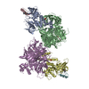
| Title | Ternary complex structure of Cereblon-DDB1 bound to WIZ(ZF7) and the molecular glue WIZ-5 | ||||||
 Components Components |
| ||||||
 Keywords Keywords | LIGASE / Cereblon / Molecular Glue | ||||||
| Function / homology |  Function and homology information Function and homology informationpositive regulation of nuclear cell cycle DNA replication / negative regulation of monoatomic ion transmembrane transport / histone methyltransferase binding / positive regulation by virus of viral protein levels in host cell / spindle assembly involved in female meiosis / epigenetic programming in the zygotic pronuclei / UV-damage excision repair / biological process involved in interaction with symbiont / regulation of mitotic cell cycle phase transition / WD40-repeat domain binding ...positive regulation of nuclear cell cycle DNA replication / negative regulation of monoatomic ion transmembrane transport / histone methyltransferase binding / positive regulation by virus of viral protein levels in host cell / spindle assembly involved in female meiosis / epigenetic programming in the zygotic pronuclei / UV-damage excision repair / biological process involved in interaction with symbiont / regulation of mitotic cell cycle phase transition / WD40-repeat domain binding / Cul4A-RING E3 ubiquitin ligase complex / Cul4-RING E3 ubiquitin ligase complex / Cul4B-RING E3 ubiquitin ligase complex / ubiquitin ligase complex scaffold activity / negative regulation of reproductive process / negative regulation of developmental process / locomotory exploration behavior / viral release from host cell / cullin family protein binding / positive regulation of Wnt signaling pathway / ectopic germ cell programmed cell death / positive regulation of viral genome replication / negative regulation of protein-containing complex assembly / proteasomal protein catabolic process / positive regulation of gluconeogenesis / transcription corepressor binding / nucleotide-excision repair / positive regulation of protein-containing complex assembly / Recognition of DNA damage by PCNA-containing replication complex / regulation of circadian rhythm / DNA Damage Recognition in GG-NER / Dual Incision in GG-NER / Transcription-Coupled Nucleotide Excision Repair (TC-NER) / Formation of TC-NER Pre-Incision Complex / Formation of Incision Complex in GG-NER / Wnt signaling pathway / Dual incision in TC-NER / Gap-filling DNA repair synthesis and ligation in TC-NER / positive regulation of protein catabolic process / cellular response to UV / rhythmic process / site of double-strand break / Neddylation / ubiquitin-dependent protein catabolic process / midbody / protein-macromolecule adaptor activity / Potential therapeutics for SARS / proteasome-mediated ubiquitin-dependent protein catabolic process / damaged DNA binding / transmembrane transporter binding / DNA-binding transcription factor activity, RNA polymerase II-specific / chromosome, telomeric region / protein stabilization / protein ubiquitination / RNA polymerase II cis-regulatory region sequence-specific DNA binding / DNA repair / apoptotic process / DNA damage response / regulation of transcription by RNA polymerase II / negative regulation of apoptotic process / protein-containing complex binding / nucleolus / perinuclear region of cytoplasm / protein-containing complex / extracellular space / DNA binding / extracellular exosome / zinc ion binding / nucleoplasm / metal ion binding / nucleus / membrane / cytosol / cytoplasm Similarity search - Function | ||||||
| Biological species |  Homo sapiens (human) Homo sapiens (human) | ||||||
| Method |  X-RAY DIFFRACTION / SYNCHROTRON / MOLECULAR REPLACEMENT / Resolution: 2.95 Å X-RAY DIFFRACTION / SYNCHROTRON / MOLECULAR REPLACEMENT / Resolution: 2.95 Å | ||||||
 Authors Authors | Partridge, J.R. / Ma, X. / Ornelas, E. | ||||||
| Funding support | 1items
| ||||||
 Citation Citation | Journal: J.Med.Chem. / Year: 2024 Title: Discovery and Optimization of First-in-Class Molecular Glue Degraders of the WIZ Transcription Factor for Fetal Hemoglobin Induction to Treat Sickle Cell Disease. Authors: Kerrigan, J.R. / Thomsen, N.M. / Cernijenko, A. / Kochanek, S.E. / Dewhurst, J. / O'Brien, G. / Ware, N.F. / Sanchez, C.C. / Manning, J.R. / Ma, X. / Ornelas, E. / Savage, N.A. / Partridge, ...Authors: Kerrigan, J.R. / Thomsen, N.M. / Cernijenko, A. / Kochanek, S.E. / Dewhurst, J. / O'Brien, G. / Ware, N.F. / Sanchez, C.C. / Manning, J.R. / Ma, X. / Ornelas, E. / Savage, N.A. / Partridge, J.R. / Patterson, A.W. / Lam, P. / Dales, N.A. / Bonazzi, S. / Borikar, S. / Hinman, A.E. / Ting, P.Y. #1: Journal: Acta Crystallogr D Struct Biol / Year: 2019 Title: Macromolecular structure determination using X-rays, neutrons and electrons: recent developments in Phenix. Authors: Dorothee Liebschner / Pavel V Afonine / Matthew L Baker / Gábor Bunkóczi / Vincent B Chen / Tristan I Croll / Bradley Hintze / Li Wei Hung / Swati Jain / Airlie J McCoy / Nigel W Moriarty ...Authors: Dorothee Liebschner / Pavel V Afonine / Matthew L Baker / Gábor Bunkóczi / Vincent B Chen / Tristan I Croll / Bradley Hintze / Li Wei Hung / Swati Jain / Airlie J McCoy / Nigel W Moriarty / Robert D Oeffner / Billy K Poon / Michael G Prisant / Randy J Read / Jane S Richardson / David C Richardson / Massimo D Sammito / Oleg V Sobolev / Duncan H Stockwell / Thomas C Terwilliger / Alexandre G Urzhumtsev / Lizbeth L Videau / Christopher J Williams / Paul D Adams /    Abstract: Diffraction (X-ray, neutron and electron) and electron cryo-microscopy are powerful methods to determine three-dimensional macromolecular structures, which are required to understand biological ...Diffraction (X-ray, neutron and electron) and electron cryo-microscopy are powerful methods to determine three-dimensional macromolecular structures, which are required to understand biological processes and to develop new therapeutics against diseases. The overall structure-solution workflow is similar for these techniques, but nuances exist because the properties of the reduced experimental data are different. Software tools for structure determination should therefore be tailored for each method. Phenix is a comprehensive software package for macromolecular structure determination that handles data from any of these techniques. Tasks performed with Phenix include data-quality assessment, map improvement, model building, the validation/rebuilding/refinement cycle and deposition. Each tool caters to the type of experimental data. The design of Phenix emphasizes the automation of procedures, where possible, to minimize repetitive and time-consuming manual tasks, while default parameters are chosen to encourage best practice. A graphical user interface provides access to many command-line features of Phenix and streamlines the transition between programs, project tracking and re-running of previous tasks. | ||||||
| History |
|
- Structure visualization
| Structure viewer | Molecule: MolmilJmol/JSmol |
|---|
- Downloads & links
-Download
| PDBx/mmCIF format | 9djt.cif.gz | 906.9 KB | Display | PDBx/mmCIF format |
|---|---|---|---|---|
| PDB format | pdb9djt.ent.gz | Display | PDB format | |
| PDBx/mmJSON format | 9djt.json.gz | Tree view | PDBx/mmJSON format | |
| Others |  Other downloads Other downloads |
-Validation report
| Arichive directory | https://data.pdbj.org/pub/pdb/validation_reports/dj/9djtftp://data.pdbj.org/pub/pdb/validation_reports/dj/9djt | HTTPS FTP |
|---|
-Related structure data

-Links
 PDBj
PDBj


- Assembly
| Deposited unit | 
| ||||||||||
|---|---|---|---|---|---|---|---|---|---|---|---|
| 1 | 
| ||||||||||
| 2 | 
| ||||||||||
| Unit cell |
|
-Components
-Protein , 2 types, 4 molecules ADBE
| #1: Protein | Mass: 42971.574 Da / Num. of mol.: 2 Source method: isolated from a genetically manipulated source Source: (gene. exp.) Homo sapiens (human) / Gene: CRBN / Production host:   Spodoptera frugiperda (fall armyworm) / References: UniProt: Q96SW2 Spodoptera frugiperda (fall armyworm) / References: UniProt: Q96SW2#2: Protein | Mass: 93347.078 Da / Num. of mol.: 2 Source method: isolated from a genetically manipulated source Source: (gene. exp.) Homo sapiens (human) / Gene: DDB1, XAP1 / Production host: Spodoptera frugiperda (fall armyworm) / References: UniProt: Q16531 |
|---|
-Protein/peptide , 1 types, 2 molecules CF
| #3: Protein/peptide | Mass: 3299.722 Da / Num. of mol.: 2 Source method: isolated from a genetically manipulated source Source: (gene. exp.) Homo sapiens (human) / Gene: WIZ, ZNF803 / Production host:  |
|---|
-Non-polymers , 4 types, 50 molecules 


| #4: Chemical | ChemComp-ZN /  Mass: 65.409 Da / Num. of mol.: 4 / Source method: obtained synthetically / Formula: Zn Mass: 65.409 Da / Num. of mol.: 4 / Source method: obtained synthetically / Formula: Zn#5: Chemical |  Mass: 96.063 Da / Num. of mol.: 2 / Source method: obtained synthetically / Formula: SO4 Mass: 96.063 Da / Num. of mol.: 2 / Source method: obtained synthetically / Formula: SO4#6: Chemical | Mass: 397.467 Da / Num. of mol.: 2 / Source method: obtained synthetically / Formula: C22H27N3O4 / Feature type: SUBJECT OF INVESTIGATION #7: Water | ChemComp-HOH / | Mass: 18.015 Da / Num. of mol.: 42 / Source method: isolated from a natural source / Formula: H2O |
|---|
-Details
| Has ligand of interest | Y |
|---|---|
| Has protein modification | Y |
-Experimental details
-Experiment
| Experiment | Method: X-RAY DIFFRACTION / Number of used crystals: 1 |
|---|
- Sample preparation
| Crystal | Density Matthews: 2.75 Å3/Da / Density % sol: 55.23 % |
|---|---|
| Crystal grow | Temperature: 291 K / Method: vapor diffusion, hanging drop / pH: 7.5 Details: 0.2 M Sodium Sulfate, 0.1 M Bis-Tris Propane pH 7.5, 20% w/v PEG 3350 |
-Data collection
| Diffraction | Mean temperature: 100 K / Serial crystal experiment: N |
|---|---|
| Diffraction source | Source: SYNCHROTRON / Site: APS / Beamline: 17-ID / Wavelength: 1 Å |
| Detector | Type: DECTRIS EIGER2 S 9M / Detector: PIXEL / Date: Apr 1, 2021 |
| Radiation | Protocol: SINGLE WAVELENGTH / Monochromatic (M) / Laue (L): M / Scattering type: x-ray |
| Radiation wavelength | Wavelength: 1 Å / Relative weight: 1 |
| Reflection | Resolution: 2.95→29.96 Å / Num. obs: 64947 / % possible obs: 99.3 % / Redundancy: 13.9 % / Biso Wilson estimate: 97.55 Å2 / CC1/2: 0.998 / Rsym value: 0.227 / Net I/σ(I): 8.9 |
| Reflection shell | Resolution: 2.95→3.02 Å / Redundancy: 14 % / Num. unique obs: 4489 / CC1/2: 0.303 / % possible all: 99 |
- Processing
| Software |
| ||||||||||||||||||||||||||||||||||||||||||||||||||||||||||||||||||||||||||||||||||||||||||||||||||||||||||||||||||||||||||||||||||||||||||||||||||||||||||||||||||||||||
|---|---|---|---|---|---|---|---|---|---|---|---|---|---|---|---|---|---|---|---|---|---|---|---|---|---|---|---|---|---|---|---|---|---|---|---|---|---|---|---|---|---|---|---|---|---|---|---|---|---|---|---|---|---|---|---|---|---|---|---|---|---|---|---|---|---|---|---|---|---|---|---|---|---|---|---|---|---|---|---|---|---|---|---|---|---|---|---|---|---|---|---|---|---|---|---|---|---|---|---|---|---|---|---|---|---|---|---|---|---|---|---|---|---|---|---|---|---|---|---|---|---|---|---|---|---|---|---|---|---|---|---|---|---|---|---|---|---|---|---|---|---|---|---|---|---|---|---|---|---|---|---|---|---|---|---|---|---|---|---|---|---|---|---|---|---|---|---|---|---|
| Refinement | Method to determine structure: MOLECULAR REPLACEMENT / Resolution: 2.95→29.96 Å / SU ML: 0.48 / Cross valid method: FREE R-VALUE / σ(F): 1.33 / Phase error: 31.59 / Stereochemistry target values: ML
| ||||||||||||||||||||||||||||||||||||||||||||||||||||||||||||||||||||||||||||||||||||||||||||||||||||||||||||||||||||||||||||||||||||||||||||||||||||||||||||||||||||||||
| Solvent computation | Shrinkage radii: 0.9 Å / VDW probe radii: 1.1 Å / Solvent model: FLAT BULK SOLVENT MODEL | ||||||||||||||||||||||||||||||||||||||||||||||||||||||||||||||||||||||||||||||||||||||||||||||||||||||||||||||||||||||||||||||||||||||||||||||||||||||||||||||||||||||||
| Refinement step | Cycle: LAST / Resolution: 2.95→29.96 Å
| ||||||||||||||||||||||||||||||||||||||||||||||||||||||||||||||||||||||||||||||||||||||||||||||||||||||||||||||||||||||||||||||||||||||||||||||||||||||||||||||||||||||||
| Refine LS restraints |
| ||||||||||||||||||||||||||||||||||||||||||||||||||||||||||||||||||||||||||||||||||||||||||||||||||||||||||||||||||||||||||||||||||||||||||||||||||||||||||||||||||||||||
| LS refinement shell |
| ||||||||||||||||||||||||||||||||||||||||||||||||||||||||||||||||||||||||||||||||||||||||||||||||||||||||||||||||||||||||||||||||||||||||||||||||||||||||||||||||||||||||
| Refinement TLS params. | Method: refined / Origin x: 27.7128 Å / Origin y: 10.8362 Å / Origin z: -20.4147 Å
| ||||||||||||||||||||||||||||||||||||||||||||||||||||||||||||||||||||||||||||||||||||||||||||||||||||||||||||||||||||||||||||||||||||||||||||||||||||||||||||||||||||||||
| Refinement TLS group | Selection details: all |
