 Movie
Movie Controller

[English] 日本語
 Yorodumi
- PDB-9dh0: The Cryo-EM structure of recombinantly expressed hUGDH in complex... -
+ Open data

- Basic information
| Entry | Database: PDB / ID: 9dh0 | ||||||||||||||||||||||||
|---|---|---|---|---|---|---|---|---|---|---|---|---|---|---|---|---|---|---|---|---|---|---|---|---|---|
| Title | The Cryo-EM structure of recombinantly expressed hUGDH in complex with UDP-4-keto-xylose | ||||||||||||||||||||||||
 Components Components | UDP-glucose 6-dehydrogenase | ||||||||||||||||||||||||
 Keywords Keywords | OXIDOREDUCTASE/INHIBITOR / Human UDP-Glucose Dehydrogenase / UDP-4-keto-xylose / OXIDOREDUCTASE / OXIDOREDUCTASE-INHIBITOR complex | ||||||||||||||||||||||||
| Function / homology |  Function and homology information Function and homology informationFormation of the active cofactor, UDP-glucuronate / UDP-glucose 6-dehydrogenase / UDP-glucose 6-dehydrogenase activity / UDP-glucuronate biosynthetic process / glycosaminoglycan biosynthetic process / chondroitin sulfate proteoglycan biosynthetic process / heparan sulfate proteoglycan biosynthetic process / gastrulation with mouth forming second / protein hexamerization / neuron development ...Formation of the active cofactor, UDP-glucuronate / UDP-glucose 6-dehydrogenase / UDP-glucose 6-dehydrogenase activity / UDP-glucuronate biosynthetic process / glycosaminoglycan biosynthetic process / chondroitin sulfate proteoglycan biosynthetic process / heparan sulfate proteoglycan biosynthetic process / gastrulation with mouth forming second / protein hexamerization / neuron development / NAD binding / extracellular exosome / nucleoplasm / identical protein binding / nucleus / cytosol Similarity search - Function | ||||||||||||||||||||||||
| Biological species |  Homo sapiens (human) Homo sapiens (human) | ||||||||||||||||||||||||
| Method | ELECTRON MICROSCOPY / single particle reconstruction / cryo EM / Resolution: 2.38 Å | ||||||||||||||||||||||||
 Authors Authors | Kadirvelraj, R. / Walsh Jr, R.M. / Wood, Z.W. | ||||||||||||||||||||||||
| Funding support |  United States, 1items United States, 1items
| ||||||||||||||||||||||||
 Citation Citation | Journal: Biochemistry / Year: 2025 Title: Cryo-EM Structure of Recombinantly Expressed hUGDH Unveils a Hidden, Alternative Allosteric Inhibitor. Authors: John H O'Brien / Renuka Kadirvelraj / Po-Sen Tseng / Nolan Ross-Kemppinen / David Crich / Richard M Walsh / Zachary A Wood / Abstract: Human UDP-glucose dehydrogenase (hUGDH) catalyzes the oxidation of UDP-glucose into UDP-glucuronic acid, an essential substrate in the Phase II metabolism of drugs. hUGDH is a hexamer that exists in ...Human UDP-glucose dehydrogenase (hUGDH) catalyzes the oxidation of UDP-glucose into UDP-glucuronic acid, an essential substrate in the Phase II metabolism of drugs. hUGDH is a hexamer that exists in an equilibrium between an active (E) state and an inactive (E) state, with the latter being stabilized by the binding of the allosteric inhibitor UDP-xylose (UDP-Xyl). The allosteric transition between E and E is slow and can be observed as a lag in progress curves. Previous analysis of the lag suggested that unliganded hUGDH exists mainly as E, but two unique crystal forms suggest that the enzyme favors the E state. Resolving this discrepancy is necessary to fully understand the allosteric mechanism of hUGDH. Here, we used cryo-EM to show that recombinant hUGDH expressed in copurifies with UDP-4-keto-xylose (UX4O), which mimics the UDP-Xyl inhibitor and favors the E state. Cryo-EM studies show that removing UX4O from hUGDH shifts the ensemble to favor the E state. This shift is consistent with progress curve analysis, which shows the absence of a lag for unliganded hUGDH. Inhibition studies show that hUGDH has similar affinities for UDP-Xyl and UX4O. The discovery that UX4O inhibits allosteric hUGDH suggests that UX4O may be the physiologically relevant inhibitor of allosteric UGDHs in bacteria that do not make UDP-Xyl. #1: Journal: Acta Crystallogr D Struct Biol / Year: 2019 Title: Macromolecular structure determination using X-rays, neutrons and electrons: recent developments in Phenix. Authors: Dorothee Liebschner / Pavel V Afonine / Matthew L Baker / Gábor Bunkóczi / Vincent B Chen / Tristan I Croll / Bradley Hintze / Li Wei Hung / Swati Jain / Airlie J McCoy / Nigel W Moriarty ...Authors: Dorothee Liebschner / Pavel V Afonine / Matthew L Baker / Gábor Bunkóczi / Vincent B Chen / Tristan I Croll / Bradley Hintze / Li Wei Hung / Swati Jain / Airlie J McCoy / Nigel W Moriarty / Robert D Oeffner / Billy K Poon / Michael G Prisant / Randy J Read / Jane S Richardson / David C Richardson / Massimo D Sammito / Oleg V Sobolev / Duncan H Stockwell / Thomas C Terwilliger / Alexandre G Urzhumtsev / Lizbeth L Videau / Christopher J Williams / Paul D Adams /   Abstract: Diffraction (X-ray, neutron and electron) and electron cryo-microscopy are powerful methods to determine three-dimensional macromolecular structures, which are required to understand biological ...Diffraction (X-ray, neutron and electron) and electron cryo-microscopy are powerful methods to determine three-dimensional macromolecular structures, which are required to understand biological processes and to develop new therapeutics against diseases. The overall structure-solution workflow is similar for these techniques, but nuances exist because the properties of the reduced experimental data are different. Software tools for structure determination should therefore be tailored for each method. Phenix is a comprehensive software package for macromolecular structure determination that handles data from any of these techniques. Tasks performed with Phenix include data-quality assessment, map improvement, model building, the validation/rebuilding/refinement cycle and deposition. Each tool caters to the type of experimental data. The design of Phenix emphasizes the automation of procedures, where possible, to minimize repetitive and time-consuming manual tasks, while default parameters are chosen to encourage best practice. A graphical user interface provides access to many command-line features of Phenix and streamlines the transition between programs, project tracking and re-running of previous tasks. | ||||||||||||||||||||||||
| History |
|
- Structure visualization
| Structure viewer | Molecule: MolmilJmol/JSmol |
|---|
- Downloads & links
-Download
| PDBx/mmCIF format | 9dh0.cif.gz | 461.9 KB | Display | PDBx/mmCIF format |
|---|---|---|---|---|
| PDB format | pdb9dh0.ent.gz | Display | PDB format | |
| PDBx/mmJSON format | 9dh0.json.gz | Tree view | PDBx/mmJSON format | |
| Others |  Other downloads Other downloads |
-Validation report
| Arichive directory | https://data.pdbj.org/pub/pdb/validation_reports/dh/9dh0ftp://data.pdbj.org/pub/pdb/validation_reports/dh/9dh0 | HTTPS FTP |
|---|
-Related structure data
| Related structure data |  46854MC 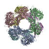 9dgzC M: map data used to model this data C: citing same article ( |
|---|---|
| Similar structure data |

-Links
 PDBj
PDBj


- Assembly
| Deposited unit | 
|
|---|---|
| 1 |
|
-Components
| #1: Protein | Mass: 55093.938 Da / Num. of mol.: 6 Source method: isolated from a genetically manipulated source Source: (gene. exp.) Homo sapiens (human) / Gene: UGDH / Production host:  #2: Chemical | ChemComp-A1BCU / ( Mass: 534.260 Da / Num. of mol.: 4 / Source method: obtained synthetically / Formula: C14H20N2O16P2 / Feature type: SUBJECT OF INVESTIGATION #3: Water | ChemComp-HOH / |  Mass: 18.015 Da / Num. of mol.: 193 / Source method: isolated from a natural source / Formula: H2O Mass: 18.015 Da / Num. of mol.: 193 / Source method: isolated from a natural source / Formula: H2OHas ligand of interest | Y | Has protein modification | N | |
|---|
-Experimental details
-Experiment
| Experiment | Method: ELECTRON MICROSCOPY |
|---|---|
| EM experiment | Aggregation state: PARTICLE / 3D reconstruction method: single particle reconstruction |
- Sample preparation
| Component | Name: Human UDP-Glucose Dehydrogenase in complex with UDP-4-keto-xylose Type: COMPLEX / Entity ID: #1 / Source: RECOMBINANT | ||||||||||||||||||||
|---|---|---|---|---|---|---|---|---|---|---|---|---|---|---|---|---|---|---|---|---|---|
| Molecular weight | Value: 0.055024 MDa / Experimental value: NO | ||||||||||||||||||||
| Source (natural) | Organism: Homo sapiens (human) | ||||||||||||||||||||
| Source (recombinant) | Organism: | ||||||||||||||||||||
| Buffer solution | pH: 7.5 | ||||||||||||||||||||
| Buffer component |
| ||||||||||||||||||||
| Specimen | Embedding applied: NO / Shadowing applied: NO / Staining applied: NO / Vitrification applied: YES / Details: 20 mM Hepes pH 7.5, 100 mM NaCl and 1 mM DTT. | ||||||||||||||||||||
| Specimen support | Grid material: GOLD / Grid mesh size: 300 divisions/in. / Grid type: Quantifoil R0.6/1 | ||||||||||||||||||||
| Vitrification | Instrument: FEI VITROBOT MARK IV / Cryogen name: ETHANE / Humidity: 100 % / Chamber temperature: 295.15 K |
- Electron microscopy imaging
| Experimental equipment |  Model: Titan Krios / Image courtesy: FEI Company |
|---|---|
| Microscopy | Model: TFS KRIOS |
| Electron gun | Electron source:  FIELD EMISSION GUN / Accelerating voltage: 300 kV / Illumination mode: FLOOD BEAM FIELD EMISSION GUN / Accelerating voltage: 300 kV / Illumination mode: FLOOD BEAM |
| Electron lens | Mode: BRIGHT FIELD / Nominal defocus max: 2000 nm / Nominal defocus min: 800 nm / C2 aperture diameter: 50 µm |
| Specimen holder | Cryogen: NITROGEN / Specimen holder model: FEI TITAN KRIOS AUTOGRID HOLDER |
| Image recording | Electron dose: 54.42 e/Å2 / Film or detector model: FEI FALCON IV (4k x 4k) / Num. of grids imaged: 1 / Num. of real images: 13500 |
- Processing
| EM software |
| ||||||||||||||||||||||||||||||||
|---|---|---|---|---|---|---|---|---|---|---|---|---|---|---|---|---|---|---|---|---|---|---|---|---|---|---|---|---|---|---|---|---|---|
| CTF correction | Type: PHASE FLIPPING AND AMPLITUDE CORRECTION | ||||||||||||||||||||||||||||||||
| Particle selection | Num. of particles selected: 4118676 | ||||||||||||||||||||||||||||||||
| Symmetry | Point symmetry: C2 (2 fold cyclic) | ||||||||||||||||||||||||||||||||
| 3D reconstruction | Resolution: 2.38 Å / Resolution method: FSC 0.143 CUT-OFF / Num. of particles: 3059917 / Symmetry type: POINT |
