 Movie
Movie Controller

[English] 日本語
 Yorodumi
- PDB-9dfs: Structure of novel Myo7a-N isoform (ADP-bound) expressed in senso... -
+ Open data

- Basic information
| Entry | Database: PDB / ID: 9dfs | ||||||||||||||||||||||||
|---|---|---|---|---|---|---|---|---|---|---|---|---|---|---|---|---|---|---|---|---|---|---|---|---|---|
| Title | Structure of novel Myo7a-N isoform (ADP-bound) expressed in sensory hair cells (head domain + first two IQ domains), bound to F-actin | ||||||||||||||||||||||||
 Components Components |
| ||||||||||||||||||||||||
 Keywords Keywords | MOTOR PROTEIN / actomyosin / hair cells | ||||||||||||||||||||||||
| Function / homology |  Function and homology information Function and homology informationmechanoreceptor differentiation / pigment granule localization / pigment granule transport / upper tip-link density / The canonical retinoid cycle in rods (twilight vision) / myosin VII complex / stereocilium base / phagolysosome assembly / inner ear receptor cell differentiation / equilibrioception ...mechanoreceptor differentiation / pigment granule localization / pigment granule transport / upper tip-link density / The canonical retinoid cycle in rods (twilight vision) / myosin VII complex / stereocilium base / phagolysosome assembly / inner ear receptor cell differentiation / equilibrioception / inner ear receptor cell stereocilium organization / sensory perception of light stimulus / photoreceptor connecting cilium / inner ear auditory receptor cell differentiation / sensory organ development / actin filament-based movement / auditory receptor cell stereocilium organization / stereocilium / inner ear morphogenesis / myosin complex / lysosome organization / microfilament motor activity / cytoskeletal motor activator activity / sensory perception / inner ear development / spectrin binding / myosin heavy chain binding / tropomyosin binding / actin filament bundle / troponin I binding / filamentous actin / microvillus / mesenchyme migration / cytoskeletal motor activity / skeletal muscle myofibril / phagocytosis / cochlea development / striated muscle thin filament / actin filament bundle assembly / skeletal muscle thin filament assembly / actin monomer binding / photoreceptor outer segment / visual perception / skeletal muscle fiber development / sensory perception of sound / actin filament polymerization / stress fiber / titin binding / photoreceptor inner segment / filopodium / actin filament / intracellular protein transport / endocytosis / ADP binding / intracellular protein localization / Hydrolases; Acting on acid anhydrides; Acting on acid anhydrides to facilitate cellular and subcellular movement / calcium-dependent protein binding / actin filament binding / actin cytoskeleton / melanosome / lamellipodium / actin binding / cell body / cell cortex / calmodulin binding / apical plasma membrane / protein domain specific binding / lysosomal membrane / positive regulation of gene expression / calcium ion binding / synapse / protein-containing complex binding / magnesium ion binding / ATP hydrolysis activity / ATP binding / identical protein binding / cytosol / cytoplasm Similarity search - Function | ||||||||||||||||||||||||
| Biological species |   Oryctolagus cuniculus (rabbit) Oryctolagus cuniculus (rabbit) Mus musculus (house mouse) Mus musculus (house mouse) | ||||||||||||||||||||||||
| Method | ELECTRON MICROSCOPY / helical reconstruction / cryo EM / Resolution: 3.1 Å | ||||||||||||||||||||||||
 Authors Authors | Egelman, E.H. / Shin, J.B. | ||||||||||||||||||||||||
| Funding support |  United States, 1items United States, 1items
| ||||||||||||||||||||||||
 Citation Citation | Journal: Nat Commun / Year: 2026 Title: Tonotopic specialization of MYO7A isoforms in auditory hair cells. Authors: Sihan Li / Jinho Park / Tobey M Phan / Giusy A Caprara / Natchanon Sittipongpittaya / Gloria M Sheynkman / Anthony W Peng / Edward H Egelman / Jonathan E Bird / Jung-Bum Shin / Abstract: Mutations in Myo7a cause Usher syndrome type 1B and non-syndromic deafness, but the precise function of MYO7A in sensory hair cells remains unclear. Using long-read sequencing, we identify and ...Mutations in Myo7a cause Usher syndrome type 1B and non-syndromic deafness, but the precise function of MYO7A in sensory hair cells remains unclear. Using long-read sequencing, we identify and characterize a novel isoform, MYO7A-N, expressed in auditory hair cells alongside the canonical MYO7A-C. Isoform-specific knock-in mouse models reveal that inner hair cells primarily express MYO7A-C, while outer hair cells express both isoforms in opposing tonotopic gradients. Both isoforms are localized to the upper tip-link insertion site, consistent with a role in the tip link for mechanotransduction. Loss of MYO7A-N leads to outer hair cell degeneration and progressive hearing loss. Cryo-EM structures reveal isoform-specific differences at actomyosin interfaces, correlating with distinct ATPase activities. These findings reveal an unexpected layer of molecular diversity within the mechanotransduction machinery. We propose that MYO7A isoform specialization enables fine-tuning of tip-link tension, thus hearing sensitivity, and contributes to the frequency-resolving power of the cochlea. | ||||||||||||||||||||||||
| History |
|
- Structure visualization
| Structure viewer | Molecule: MolmilJmol/JSmol |
|---|
- Downloads & links
-Download
| PDBx/mmCIF format | 9dfs.cif.gz | 372.1 KB | Display | PDBx/mmCIF format |
|---|---|---|---|---|
| PDB format | pdb9dfs.ent.gz | 292.7 KB | Display | PDB format |
| PDBx/mmJSON format | 9dfs.json.gz | Tree view | PDBx/mmJSON format | |
| Others |  Other downloads Other downloads |
-Validation report
| Arichive directory | https://data.pdbj.org/pub/pdb/validation_reports/df/9dfsftp://data.pdbj.org/pub/pdb/validation_reports/df/9dfs | HTTPS FTP |
|---|
-Related structure data
| Related structure data |  46818MC  9dfvC M: map data used to model this data C: citing same article ( |
|---|---|
| Similar structure data |

-Links
 PDBj
PDBj

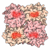

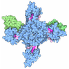


- Assembly
| Deposited unit | 
|
|---|---|
| 1 |
|
-Components
| #1: Protein | Mass: 42109.973 Da / Num. of mol.: 3 Source method: isolated from a genetically manipulated source Source: (gene. exp.) Oryctolagus cuniculus (rabbit) / Tissue: skeletal muscle / Gene: ACTA1, ACTA / Production host: Oryctolagus cuniculus (rabbit)References: UniProt: P68135, Hydrolases; Acting on acid anhydrides; Acting on acid anhydrides to facilitate cellular and subcellular movement #2: Protein | | Mass: 122808.883 Da / Num. of mol.: 1 Source method: isolated from a genetically manipulated source Details: Myo7a-N isoform (ADP-bound) expressed in sensory hair cells (head domain + first two IQ domains) Source: (gene. exp.) Mus musculus (house mouse) / Gene: Myo7a, Myo7 / Production host:  unidentified baculovirus / References: UniProt: P97479 unidentified baculovirus / References: UniProt: P97479#3: Chemical | ChemComp-ADP /  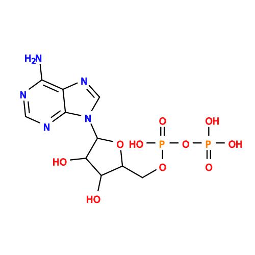 Mass: 427.201 Da / Num. of mol.: 4 / Source method: obtained synthetically / Formula: C10H15N5O10P2 / Comment: ADP, energy-carrying molecule*YM Mass: 427.201 Da / Num. of mol.: 4 / Source method: obtained synthetically / Formula: C10H15N5O10P2 / Comment: ADP, energy-carrying molecule*YM#4: Chemical | ChemComp-MG /   Mass: 24.305 Da / Num. of mol.: 4 / Source method: obtained synthetically / Formula: Mg Mass: 24.305 Da / Num. of mol.: 4 / Source method: obtained synthetically / Formula: MgHas ligand of interest | N | Has protein modification | Y | |
|---|
-Experimental details
-Experiment
| Experiment | Method: ELECTRON MICROSCOPY |
|---|---|
| EM experiment | Aggregation state: FILAMENT / 3D reconstruction method: helical reconstruction |
- Sample preparation
| Component | Name: actomyosin / Type: COMPLEX / Entity ID: #1-#2 / Source: NATURAL |
|---|---|
| Molecular weight | Experimental value: NO |
| Source (natural) | Organism: Oryctolagus cuniculus (rabbit) |
| Buffer solution | pH: 7.8 |
| Specimen | Embedding applied: NO / Shadowing applied: NO / Staining applied: NO / Vitrification applied: YES |
| Vitrification | Cryogen name: ETHANE |
- Electron microscopy imaging
| Experimental equipment |  Model: Titan Krios / Image courtesy: FEI Company |
|---|---|
| Microscopy | Model: FEI TITAN KRIOS |
| Electron gun | Electron source:  FIELD EMISSION GUN / Accelerating voltage: 300 kV / Illumination mode: FLOOD BEAM FIELD EMISSION GUN / Accelerating voltage: 300 kV / Illumination mode: FLOOD BEAM |
| Electron lens | Mode: BRIGHT FIELD / Nominal defocus max: 2500 nm / Nominal defocus min: 600 nm |
| Image recording | Electron dose: 50 e/Å2 / Film or detector model: GATAN K3 BIOQUANTUM (6k x 4k) |
- Processing
| EM software | Name: PHENIX / Version: 1.20.1_4487: / Category: model refinement | ||||||||||||||||||||||||
|---|---|---|---|---|---|---|---|---|---|---|---|---|---|---|---|---|---|---|---|---|---|---|---|---|---|
| CTF correction | Type: PHASE FLIPPING AND AMPLITUDE CORRECTION | ||||||||||||||||||||||||
| Helical symmerty | Angular rotation/subunit: 0 ° / Axial rise/subunit: 0.001 Å / Axial symmetry: C1 | ||||||||||||||||||||||||
| 3D reconstruction | Resolution: 3.1 Å / Resolution method: FSC 0.143 CUT-OFF / Num. of particles: 878646 / Symmetry type: HELICAL | ||||||||||||||||||||||||
| Refine LS restraints |
|
