 Movie
Movie Controller


+ Open data

- Basic information
| Entry | Database: PDB / ID: 9dcb | ||||||
|---|---|---|---|---|---|---|---|
| Title | The Structure of AAV5 at 4 Degrees | ||||||
 Components Components |
| ||||||
 Keywords Keywords | VIRUS / temperature / genome / vector / icosahedron | ||||||
| Function / homology |  Function and homology information Function and homology information | ||||||
| Biological species |  adeno-associated virus 5 adeno-associated virus 5 | ||||||
| Method | ELECTRON MICROSCOPY / single particle reconstruction / cryo EM / Resolution: 2.89 Å | ||||||
 Authors Authors | Bennett, A.B. / McKenna, R. | ||||||
| Funding support |  United States, 1items United States, 1items
| ||||||
 Citation Citation | Journal: J Virol / Year: 2025 Title: Biophysical and structural insights into AAV genome ejection. Authors: Keely Gliwa / Joshua Hull / Austin Kansol / Victoria Zembruski / Renuk Lakshmanan / Mario Mietzsch / Paul Chipman / Antonette Bennett / Robert McKenna / Abstract: Recombinant adeno-associated virus (rAAV) is comprised of non-enveloped capsids that can package a therapeutic transgene and are currently being developed and utilized as gene therapy vectors. The ...Recombinant adeno-associated virus (rAAV) is comprised of non-enveloped capsids that can package a therapeutic transgene and are currently being developed and utilized as gene therapy vectors. The therapeutic efficiency of rAAV is dependent on successful cytoplasmic trafficking and transgene delivery to the nucleus. It is hypothesized that an increased understanding of the effects of the cellular environment and biophysical properties of the capsid as it traffics to the nucleus could provide insight to improve vector efficiency. The AAV capsid is exposed to increasing [H] during endo-lysosomal trafficking. Exposure to low pH facilitates the externalization of the viral protein 1 unique region (VP1u). This VP1u contains a phospholipase A2 domain required for endosomal escape and nuclear localization signals that facilitate nuclear targeting and entry. The viral genome is released either after total capsid disassembly or via a concerted DNA ejection mechanism in the nucleus. This study presents the characterization of genome ejection (GE) for two diverse serotypes, AAV2 and AAV5, using temperature. The temperature required to disassemble the virus capsid (T) is significantly higher than the temperature required to expose the transgene (T) for both serotypes. This was verified by quantitative PCR (qPCR) and transmission electron microscopy. Additionally, the absence of VP1/VP2 in the capsids and a decrease in pH increase the temperature of GE. Furthermore, cryo-electron microscopy structures of the AAV5 capsid pre- and post-GE reveal dynamics at the twofold, threefold, and fivefold regions of the capsid interior consistent with a concerted egress of the viral genome.IMPORTANCEThe development of recombinant adeno-associated virus (rAAV) capsids has grown rapidly in recent years, with five of the eight established therapeutics gaining approval in the past 2 years alone. Clinical progression with AAV2 and AAV5 represents a growing need to further characterize the molecular biology of these viruses. The goal of AAV-based gene therapy is to treat monogenic disorders with a vector-delivered transgene to provide wild-type protein function. A better understanding of the dynamics and conditions enabling transgene release may improve therapeutic efficiency. In addition to their clinical importance, AAV2 and 5 were chosen in this study for their diverse antigenic and biophysical properties compared to more closely related serotypes. Characterization of a shared genome ejection process may imply a conserved mechanism for all rAAV therapies. | ||||||
| History |
|
- Structure visualization
| Structure viewer | Molecule: MolmilJmol/JSmol |
|---|
- Downloads & links
-Download
| PDBx/mmCIF format | 9dcb.cif.gz | 5.8 MB | Display | PDBx/mmCIF format |
|---|---|---|---|---|
| PDB format | pdb9dcb.ent.gz | Display | PDB format | |
| PDBx/mmJSON format | 9dcb.json.gz | Tree view | PDBx/mmJSON format | |
| Others |  Other downloads Other downloads |
-Validation report
| Arichive directory | https://data.pdbj.org/pub/pdb/validation_reports/dc/9dcbftp://data.pdbj.org/pub/pdb/validation_reports/dc/9dcb | HTTPS FTP |
|---|
-Related structure data
| Related structure data | 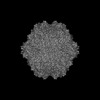 46748MC  9dc7C 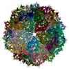 9dccC M: map data used to model this data C: citing same article ( |
|---|---|
| Similar structure data |

-Links
 PDBj
PDBj


- Assembly
| Deposited unit | 
|
|---|---|
| 1 |
|
-Components
| #1: Protein | Mass: 80497.414 Da / Num. of mol.: 60 Source method: isolated from a genetically manipulated source Source: (gene. exp.) adeno-associated virus 5 / Gene: cap, VP1 / Production host:   Spodoptera frugiperda (fall armyworm) / References: UniProt: Q9YIJ1 Spodoptera frugiperda (fall armyworm) / References: UniProt: Q9YIJ1#2: DNA chain | Mass: 581.456 Da / Num. of mol.: 60 Source method: isolated from a genetically manipulated source Source: (gene. exp.) adeno-associated virus 5 / Production host: Spodoptera frugiperda (fall armyworm)Has ligand of interest | N | Has protein modification | N | |
|---|
-Experimental details
-Experiment
| Experiment | Method: ELECTRON MICROSCOPY |
|---|---|
| EM experiment | Aggregation state: PARTICLE / 3D reconstruction method: single particle reconstruction |
- Sample preparation
| Component | Name: adeno-associated virus 5 / Type: VIRUS / Details: Baculovirus expression / Entity ID: all / Source: RECOMBINANT |
|---|---|
| Molecular weight | Value: 4 MDa / Experimental value: NO |
| Source (natural) | Organism: adeno-associated virus 5 |
| Source (recombinant) | Organism: Spodoptera frugiperda (fall armyworm) |
| Details of virus | Empty: NO / Enveloped: NO / Isolate: SEROTYPE / Type: VIRION |
| Buffer solution | pH: 7.4 Details: 10 mM Na2HPO4, 2 mM KH2PO4, 135 mM NaCl, 5 mM KCl, 1 mM MgCl2, pH 7.4 |
| Buffer component | Conc.: 1 mM / Name: TD / Formula: PBS-MK |
| Specimen | Conc.: 0.5 mg/ml / Embedding applied: NO / Shadowing applied: NO / Staining applied: NO / Vitrification applied: YES |
| Vitrification | Instrument: FEI VITROBOT MARK IV / Cryogen name: ETHANE / Humidity: 95 % |
- Electron microscopy imaging
| Experimental equipment |  Model: Titan Krios / Image courtesy: FEI Company |
|---|---|
| Microscopy | Model: TFS KRIOS |
| Electron gun | Electron source:  FIELD EMISSION GUN / Accelerating voltage: 300 kV / Illumination mode: FLOOD BEAM FIELD EMISSION GUN / Accelerating voltage: 300 kV / Illumination mode: FLOOD BEAM |
| Electron lens | Mode: BRIGHT FIELD / Nominal defocus max: 3967 nm / Nominal defocus min: 1289 nm |
| Image recording | Electron dose: 45 e/Å2 / Film or detector model: FEI FALCON IV (4k x 4k) |
- Processing
| EM software | Name: PHENIX / Version: 1.10-2155_2155: / Category: model refinement | ||||||||||||||||||||||||
|---|---|---|---|---|---|---|---|---|---|---|---|---|---|---|---|---|---|---|---|---|---|---|---|---|---|
| CTF correction | Type: PHASE FLIPPING AND AMPLITUDE CORRECTION | ||||||||||||||||||||||||
| 3D reconstruction | Resolution: 2.89 Å / Resolution method: FSC 0.143 CUT-OFF / Num. of particles: 23000 / Symmetry type: POINT | ||||||||||||||||||||||||
| Refine LS restraints |
|
