 Movie
Movie Controller

[English] 日本語
 Yorodumi
- PDB-9d9u: Rhodospirillum rubrum Nitrogenase-like Methylthio-alkane Reductas... -
+ Open data

- Basic information
| Entry | Database: PDB / ID: 9d9u | ||||||||||||
|---|---|---|---|---|---|---|---|---|---|---|---|---|---|
| Title | Rhodospirillum rubrum Nitrogenase-like Methylthio-alkane Reductase Complex with an Oxidized P-cluster | ||||||||||||
 Components Components | (Nitrogenase) x 2 | ||||||||||||
 Keywords Keywords | OXIDOREDUCTASE / Methylthio-alkane reductase / Carbon-Sulfur lyase / dimethylsulfide / methane / methanethiol | ||||||||||||
| Function / homology | Oxidoreductases; Acting on iron-sulfur proteins as donors / : / : / Nitrogenase/oxidoreductase, component 1 / Nitrogenase component 1 type Oxidoreductase / nitrogenase activity / FE(8)-S(7) CLUSTER / Methylthio-alkane reductase catalytic subunit alpha / Methylthio-alkane reductase catalytic subunit beta Function and homology information Function and homology information | ||||||||||||
| Biological species |  Rhodospirillum rubrum ATCC 11170 (bacteria) Rhodospirillum rubrum ATCC 11170 (bacteria) | ||||||||||||
| Method | ELECTRON MICROSCOPY / single particle reconstruction / cryo EM / Resolution: 2.35 Å | ||||||||||||
 Authors Authors | Kreitler, D.F. / Hu, G. / North, J.A. | ||||||||||||
| Funding support |  United States, 3items United States, 3items
| ||||||||||||
 Citation Citation | Journal: Nat Catal / Year: 2025 Title: Architecture, catalysis and regulation of methylthio-alkane reductase for bacterial sulfur acquisition from volatile organic compounds Authors: Murali, S. / Hu, G.B. / Kreitler, D.F. / Carriedo, A.A. / Lewis, L.C. / Fosu, S.A. / Weaver, O.G. / Buzas, E.M. / Byerly, K.M. / Yoshikuni, Y. / McSweeney, S. / Shafaat, H.S. / North, J.A. | ||||||||||||
| History |
|
- Structure visualization
| Structure viewer | Molecule: MolmilJmol/JSmol |
|---|
- Downloads & links
-Download
| PDBx/mmCIF format | 9d9u.cif.gz | 594.7 KB | Display | PDBx/mmCIF format |
|---|---|---|---|---|
| PDB format | pdb9d9u.ent.gz | 493.4 KB | Display | PDB format |
| PDBx/mmJSON format | 9d9u.json.gz | Tree view | PDBx/mmJSON format | |
| Others |  Other downloads Other downloads |
-Validation report
| Arichive directory | https://data.pdbj.org/pub/pdb/validation_reports/d9/9d9uftp://data.pdbj.org/pub/pdb/validation_reports/d9/9d9u | HTTPS FTP |
|---|
-Related structure data
| Related structure data |  46680MC M: map data used to model this data C: citing same article ( |
|---|---|
| Similar structure data |
-Links
 PDBj
PDBj
- Assembly
| Deposited unit | 
|
|---|---|
| 1 |
|
-Components
| #1: Protein | Mass: 57613.164 Da / Num. of mol.: 2 Source method: isolated from a genetically manipulated source Source: (gene. exp.) Rhodospirillum rubrum ATCC 11170 (bacteria)Gene: Rru_A0794 Production host: Rhodospirillum rubrum ATCC 11170 (bacteria)References: UniProt: Q2RW97, nitrogenase #2: Protein | Mass: 50856.289 Da / Num. of mol.: 2 Source method: isolated from a genetically manipulated source Source: (gene. exp.) Rhodospirillum rubrum ATCC 11170 (bacteria)Gene: Rru_A0793 Production host: Rhodospirillum rubrum ATCC 11170 (bacteria)References: UniProt: Q2RW98, nitrogenase #3: Chemical | 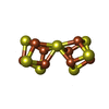  Mass: 671.215 Da / Num. of mol.: 2 / Source method: obtained synthetically / Formula: Fe8S7 / Feature type: SUBJECT OF INVESTIGATION Mass: 671.215 Da / Num. of mol.: 2 / Source method: obtained synthetically / Formula: Fe8S7 / Feature type: SUBJECT OF INVESTIGATIONHas ligand of interest | Y | Has protein modification | N | |
|---|
-Experimental details
-Experiment
| Experiment | Method: ELECTRON MICROSCOPY |
|---|---|
| EM experiment | Aggregation state: PARTICLE / 3D reconstruction method: single particle reconstruction |
- Sample preparation
| Component | Name: Complex of marDK heterotetramer under dithionite reduced conditions Type: COMPLEX / Entity ID: #1-#2 / Source: RECOMBINANT | ||||||||||||||||||||
|---|---|---|---|---|---|---|---|---|---|---|---|---|---|---|---|---|---|---|---|---|---|
| Molecular weight | Value: 0.228 MDa / Experimental value: YES | ||||||||||||||||||||
| Source (natural) | Organism: Rhodospirillum rubrum ATCC 11170 (bacteria) | ||||||||||||||||||||
| Source (recombinant) | Organism: Rhodospirillum rubrum ATCC 11170 (bacteria) / Strain: marBHDK deletion strain | ||||||||||||||||||||
| Buffer solution | pH: 7.4 | ||||||||||||||||||||
| Buffer component |
| ||||||||||||||||||||
| Specimen | Conc.: 1.9 mg/ml / Embedding applied: NO / Shadowing applied: NO / Staining applied: NO / Vitrification applied: YES Details: The sample was monodisperse, diluted with buffer from 7.5 mg/mL stock solution. | ||||||||||||||||||||
| Specimen support | Details: 30 mA / Grid material: GOLD / Grid mesh size: 300 divisions/in. / Grid type: Quantifoil R1.2/1.3 | ||||||||||||||||||||
| Vitrification | Instrument: FEI VITROBOT MARK IV / Cryogen name: ETHANE / Humidity: 100 % / Chamber temperature: 277 K Details: 3.5ul sample was applied to a glow discharged UltrAuFoil grid, blotted for 3 or 4 seconds before plunged in liquid ethane cooled with liquid nitrogen. |
- Electron microscopy imaging
| Experimental equipment |  Model: Titan Krios / Image courtesy: FEI Company |
|---|---|
| Microscopy | Model: TFS KRIOS Details: Sample was screened using negative staining and Cryo-EM on a screening microscope for sample readiness assessment before automated high-throughput and high-resolution data collection on ...Details: Sample was screened using negative staining and Cryo-EM on a screening microscope for sample readiness assessment before automated high-throughput and high-resolution data collection on Titan Krios. Multiple batches of Cryo-EM specimen were prepared in order to deal with the orientation preference. The calibrated minimum/maximum defocus were the calculated minimum and maximum of images that with contaminants or broken ice. |
| Electron gun | Electron source:  FIELD EMISSION GUN / Accelerating voltage: 300 kV / Illumination mode: FLOOD BEAM FIELD EMISSION GUN / Accelerating voltage: 300 kV / Illumination mode: FLOOD BEAM |
| Electron lens | Mode: BRIGHT FIELD / Nominal magnification: 105000 X / Calibrated magnification: 109454 X / Nominal defocus max: 2000 nm / Nominal defocus min: 1000 nm / Calibrated defocus min: 214 nm / Calibrated defocus max: 3010 nm / Cs: 2.7 mm / C2 aperture diameter: 70 µm / Alignment procedure: COMA FREE |
| Specimen holder | Cryogen: NITROGEN / Specimen holder model: FEI TITAN KRIOS AUTOGRID HOLDER / Temperature (max): 79.8 K / Temperature (min): 79.8 K |
| Image recording | Average exposure time: 2.26 sec. / Electron dose: 50 e/Å2 / Film or detector model: GATAN K3 BIOQUANTUM (6k x 4k) / Num. of grids imaged: 1 / Num. of real images: 4840 Details: Data was collected on a ThermoScientific Titan Krios G3i Cryo-TEM operated at 300kV and at a nominal 105,000 magnification using ThermoScientific EPU program with the defocus range of -1.0 ...Details: Data was collected on a ThermoScientific Titan Krios G3i Cryo-TEM operated at 300kV and at a nominal 105,000 magnification using ThermoScientific EPU program with the defocus range of -1.0 to -2.0 um . Image stack files were filtered with a BioQuantum energy filter at 15eV slit width and acquired with a Gatan K3 Direct Electron Detector and (Gatan, Pleasanton, CA, USA) in COunted Super-Resolution mode resulting pixel size 0.4125 angstrom. Total electron dose was 50 electrons. And each stack file consists of 40 frames. |
| EM imaging optics | Energyfilter name: GIF Bioquantum / Chromatic aberration corrector: N/A. / Energyfilter slit width: 15 eV / Spherical aberration corrector: N/A. |
| Image scans | Sampling size: 5 µm / Width: 5760 / Height: 4092 |
- Processing
| EM software |
| |||||||||||||||||||||||||||||||||||||||||||||
|---|---|---|---|---|---|---|---|---|---|---|---|---|---|---|---|---|---|---|---|---|---|---|---|---|---|---|---|---|---|---|---|---|---|---|---|---|---|---|---|---|---|---|---|---|---|---|
| CTF correction | Type: PHASE FLIPPING AND AMPLITUDE CORRECTION | |||||||||||||||||||||||||||||||||||||||||||||
| Particle selection | Num. of particles selected: 6195342 | |||||||||||||||||||||||||||||||||||||||||||||
| Symmetry | Point symmetry: C2 (2 fold cyclic) | |||||||||||||||||||||||||||||||||||||||||||||
| 3D reconstruction | Resolution: 2.35 Å / Resolution method: FSC 0.143 CUT-OFF / Num. of particles: 149015 / Algorithm: SIMULTANEOUS ITERATIVE (SIRT) Details: Final step was actually Non-uniform Refinement of two classes of an Ab Initio reconstruction. Num. of class averages: 2 / Symmetry type: POINT | |||||||||||||||||||||||||||||||||||||||||||||
| Atomic model building | Protocol: AB INITIO MODEL / Space: REAL | |||||||||||||||||||||||||||||||||||||||||||||
| Atomic model building | Details: AlphaFold multimer / Source name: AlphaFold / Type: in silico model | |||||||||||||||||||||||||||||||||||||||||||||
| Refinement | Highest resolution: 2.35 Å / Cross valid method: NONE Stereochemistry target values: REAL-SPACE (WEIGHTED MAP SUM AT ATOM CENTERS) |
