[English] 日本語
 Yorodumi
- EMDB-46680: Rhodospirillum rubrum Nitrogenase-like Methylthio-alkane Reductas... -
+ Open data
- Basic information
| Entry |  | ||||||||||||
|---|---|---|---|---|---|---|---|---|---|---|---|---|---|
| Title | Rhodospirillum rubrum Nitrogenase-like Methylthio-alkane Reductase Complex with an Oxidized P-cluster | ||||||||||||
 Map data Map data | |||||||||||||
 Sample Sample |
| ||||||||||||
 Keywords Keywords | Methylthio-alkane reductase / Carbon-Sulfur lyase / dimethylsulfide / methane / methanethiol / OXIDOREDUCTASE | ||||||||||||
| Function / homology | : / : / Nitrogenase/oxidoreductase, component 1 / Nitrogenase component 1 type Oxidoreductase / nitrogenase / nitrogenase activity / Nitrogenase / Nitrogenase Function and homology information Function and homology information | ||||||||||||
| Biological species |  Rhodospirillum rubrum ATCC 11170 (bacteria) Rhodospirillum rubrum ATCC 11170 (bacteria) | ||||||||||||
| Method | single particle reconstruction / cryo EM / Resolution: 2.35 Å | ||||||||||||
 Authors Authors | Kreitler DF / Hu G / North JA | ||||||||||||
| Funding support |  United States, 3 items United States, 3 items
| ||||||||||||
 Citation Citation |  Journal: Nat Catal / Year: 2025 Journal: Nat Catal / Year: 2025Title: Architecture, catalysis and regulation of methylthio-alkane reductase for bacterial sulfur acquisition from volatile organic compounds Authors: Murali S / Hu GB / Kreitler DF / Carriedo AA / Lewis LC / Fosu SA / Weaver OG / Buzas EM / Byerly KM / Yoshikuni Y / McSweeney S / Shafaat HS / North JA | ||||||||||||
| History |
|
- Structure visualization
| Supplemental images |
|---|
- Downloads & links
-EMDB archive
| Map data |  emd_46680.map.gz emd_46680.map.gz | 123 MB |  EMDB map data format EMDB map data format | |
|---|---|---|---|---|
| Header (meta data) |  emd-46680-v30.xml emd-46680-v30.xml emd-46680.xml emd-46680.xml | 24.4 KB 24.4 KB | Display Display |  EMDB header EMDB header |
| Images |  emd_46680.png emd_46680.png | 83.3 KB | ||
| Filedesc metadata |  emd-46680.cif.gz emd-46680.cif.gz | 8 KB | ||
| Others |  emd_46680_half_map_1.map.gz emd_46680_half_map_1.map.gz emd_46680_half_map_2.map.gz emd_46680_half_map_2.map.gz | 226.6 MB 226.6 MB | ||
| Archive directory |  http://ftp.pdbj.org/pub/emdb/structures/EMD-46680 http://ftp.pdbj.org/pub/emdb/structures/EMD-46680 ftp://ftp.pdbj.org/pub/emdb/structures/EMD-46680 ftp://ftp.pdbj.org/pub/emdb/structures/EMD-46680 | HTTPS FTP |
-Validation report
| Summary document |  emd_46680_validation.pdf.gz emd_46680_validation.pdf.gz | 877.3 KB | Display |  EMDB validaton report EMDB validaton report |
|---|---|---|---|---|
| Full document |  emd_46680_full_validation.pdf.gz emd_46680_full_validation.pdf.gz | 876.9 KB | Display | |
| Data in XML |  emd_46680_validation.xml.gz emd_46680_validation.xml.gz | 15.6 KB | Display | |
| Data in CIF |  emd_46680_validation.cif.gz emd_46680_validation.cif.gz | 18.4 KB | Display | |
| Arichive directory |  https://ftp.pdbj.org/pub/emdb/validation_reports/EMD-46680 https://ftp.pdbj.org/pub/emdb/validation_reports/EMD-46680 ftp://ftp.pdbj.org/pub/emdb/validation_reports/EMD-46680 ftp://ftp.pdbj.org/pub/emdb/validation_reports/EMD-46680 | HTTPS FTP |
-Related structure data
| Related structure data |  9d9uMC M: atomic model generated by this map C: citing same article ( |
|---|---|
| Similar structure data | Similarity search - Function & homology  F&H Search F&H Search |
- Links
Links
| EMDB pages |  EMDB (EBI/PDBe) / EMDB (EBI/PDBe) /  EMDataResource EMDataResource |
|---|---|
| Related items in Molecule of the Month |
- Map
Map
| File |  Download / File: emd_46680.map.gz / Format: CCP4 / Size: 244.1 MB / Type: IMAGE STORED AS FLOATING POINT NUMBER (4 BYTES) Download / File: emd_46680.map.gz / Format: CCP4 / Size: 244.1 MB / Type: IMAGE STORED AS FLOATING POINT NUMBER (4 BYTES) | ||||||||||||||||||||||||||||||||||||
|---|---|---|---|---|---|---|---|---|---|---|---|---|---|---|---|---|---|---|---|---|---|---|---|---|---|---|---|---|---|---|---|---|---|---|---|---|---|
| Projections & slices | Image control
Images are generated by Spider. | ||||||||||||||||||||||||||||||||||||
| Voxel size | X=Y=Z: 0.825 Å | ||||||||||||||||||||||||||||||||||||
| Density |
| ||||||||||||||||||||||||||||||||||||
| Symmetry | Space group: 1 | ||||||||||||||||||||||||||||||||||||
| Details | EMDB XML:
|
-Supplemental data
-Half map: #2
| File | emd_46680_half_map_1.map | ||||||||||||
|---|---|---|---|---|---|---|---|---|---|---|---|---|---|
| Projections & Slices |
| ||||||||||||
| Density Histograms |
-Half map: #1
| File | emd_46680_half_map_2.map | ||||||||||||
|---|---|---|---|---|---|---|---|---|---|---|---|---|---|
| Projections & Slices |
| ||||||||||||
| Density Histograms |
- Sample components
-Entire : Complex of marDK heterotetramer under dithionite reduced conditions
| Entire | Name: Complex of marDK heterotetramer under dithionite reduced conditions |
|---|---|
| Components |
|
-Supramolecule #1: Complex of marDK heterotetramer under dithionite reduced conditions
| Supramolecule | Name: Complex of marDK heterotetramer under dithionite reduced conditions type: complex / ID: 1 / Parent: 0 / Macromolecule list: #1-#2 |
|---|---|
| Source (natural) | Organism:  Rhodospirillum rubrum ATCC 11170 (bacteria) Rhodospirillum rubrum ATCC 11170 (bacteria) |
| Molecular weight | Theoretical: 228 KDa |
-Macromolecule #1: Nitrogenase
| Macromolecule | Name: Nitrogenase / type: protein_or_peptide / ID: 1 / Number of copies: 2 / Enantiomer: LEVO / EC number: nitrogenase |
|---|---|
| Source (natural) | Organism:  Rhodospirillum rubrum ATCC 11170 (bacteria) Rhodospirillum rubrum ATCC 11170 (bacteria) |
| Molecular weight | Theoretical: 57.613164 KDa |
| Recombinant expression | Organism:  Rhodospirillum rubrum ATCC 11170 (bacteria) Rhodospirillum rubrum ATCC 11170 (bacteria) |
| Sequence | String: MPIHHHHHHQ HNLKTSVVES REQRLGTIIA WDGKASDLSK ESAYARSEGC GSACGAKARR VCEMRSPFSQ GSVCSEQMVE CQAGNVRGA VLVQHSPIGC GAGQVIYNSI FRNGLAIRGL PVENLHLIST NLRERDMVYG GLDKLERTIR DAWERHHPQA I FIATSCPT ...String: MPIHHHHHHQ HNLKTSVVES REQRLGTIIA WDGKASDLSK ESAYARSEGC GSACGAKARR VCEMRSPFSQ GSVCSEQMVE CQAGNVRGA VLVQHSPIGC GAGQVIYNSI FRNGLAIRGL PVENLHLIST NLRERDMVYG GLDKLERTIR DAWERHHPQA I FIATSCPT AIIGDDIESV ASQLEAEFGI PVIPLHCEGF KSKHWSTGFD ATQHGILRQI VRKNPERKQE DLVNVINLWG SD VFGPMLG ELGLRVNYVV DLATVEDLAQ MSEAAATVGF CYTLSTYMAA ALEQEFGVPE VKAPMPYGFA GTDAWLREIA RVT HREEQA EAYIAREHAR VKPQLEALRE KLKGIKGFVS TGSAYAHGMI QVLRELGVTV DGSLVFHHDP VYDSQDPRQD SLAH LVDNY GDVGHFSVGN RQQFQFYGLL QRVKPDFIII RHNGLAPLAS RLGIPAIPLG DEHIAVGYQG ILNLGESILD VLAHR KFHE DIAAHVRLPY RQDWLARDPF DLARQSAGQP RRPAE UniProtKB: Nitrogenase |
-Macromolecule #2: Nitrogenase
| Macromolecule | Name: Nitrogenase / type: protein_or_peptide / ID: 2 / Number of copies: 2 / Enantiomer: LEVO / EC number: nitrogenase |
|---|---|
| Source (natural) | Organism:  Rhodospirillum rubrum ATCC 11170 (bacteria) Rhodospirillum rubrum ATCC 11170 (bacteria) |
| Molecular weight | Theoretical: 50.856289 KDa |
| Recombinant expression | Organism:  Rhodospirillum rubrum ATCC 11170 (bacteria) Rhodospirillum rubrum ATCC 11170 (bacteria) |
| Sequence | String: MPDAESRSQV TAKAAPPPAP KTNSIEQVRY ICSIGAMHSA SAIPRVIPIT HCGPGCADKQ FMNVAFYNGF QGGGYGGGAV VPSTNATER EVVFGGAERL DELIGASLQV LDADLFVVLT GCIPDLVGDD IGSVVGPYQK RGVPIVYAET GGFRGNNFTG H ELVTKAII ...String: MPDAESRSQV TAKAAPPPAP KTNSIEQVRY ICSIGAMHSA SAIPRVIPIT HCGPGCADKQ FMNVAFYNGF QGGGYGGGAV VPSTNATER EVVFGGAERL DELIGASLQV LDADLFVVLT GCIPDLVGDD IGSVVGPYQK RGVPIVYAET GGFRGNNFTG H ELVTKAII DQFVGDYDAE RDGAREPHTV NVWSLLPYHN TFWRGDLTEI KRLLEGIGLK VNILFGPQSA GVAEWKAIPR AG FNLVLSP WLGLDTARHL DRKYGQPTLH RPIIPIGAKE TGAFLREVAA FAGLDSAVVE AFITAEEAVY YRYLEDFTDF YAE YWWGLP AKFAVIGDSA YNLALTKFLV NQLGLIPGLQ IITDNPPEEV REDIRAHYHA IADDVATDVS FEEDSYTIHQ KIRA TDFGH KAPILFGTTW ERDLAKELKG AIVEVGFPAS YEVVLSRSYL GYRGALTLLE KIYTTTVSAS A UniProtKB: Nitrogenase |
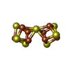
-Macromolecule #3: FE(8)-S(7) CLUSTER
| Macromolecule | Name: FE(8)-S(7) CLUSTER / type: ligand / ID: 3 / Number of copies: 2 / Formula: CLF |
|---|---|
| Molecular weight | Theoretical: 671.215 Da |
| Chemical component information |  ChemComp-CLF: |
-Experimental details
-Structure determination
| Method | cryo EM |
|---|---|
 Processing Processing | single particle reconstruction |
| Aggregation state | particle |
- Sample preparation
| Concentration | 1.9 mg/mL | ||||||||||||
|---|---|---|---|---|---|---|---|---|---|---|---|---|---|
| Buffer | pH: 7.4 Component:
| ||||||||||||
| Grid | Model: Quantifoil R1.2/1.3 / Material: GOLD / Mesh: 300 / Support film - Material: GOLD / Support film - topology: HOLEY / Support film - Film thickness: 50 / Pretreatment - Type: GLOW DISCHARGE / Pretreatment - Time: 120 sec. / Pretreatment - Pressure: 4.0000000000000003e-07 kPa / Details: 30 mA | ||||||||||||
| Vitrification | Cryogen name: ETHANE / Chamber humidity: 100 % / Chamber temperature: 277 K / Instrument: FEI VITROBOT MARK IV Details: 3.5ul sample was applied to a glow discharged UltrAuFoil grid, blotted for 3 or 4 seconds before plunged in liquid ethane cooled with liquid nitrogen.. | ||||||||||||
| Details | The sample was monodisperse, diluted with buffer from 7.5 mg/mL stock solution. |
- Electron microscopy
| Microscope | TFS KRIOS |
|---|---|
| Temperature | Min: 79.8 K / Max: 79.8 K |
| Specialist optics | Spherical aberration corrector: N/A. / Chromatic aberration corrector: N/A. / Energy filter - Name: GIF Bioquantum / Energy filter - Slit width: 15 eV |
| Details | Sample was screened using negative staining and Cryo-EM on a screening microscope for sample readiness assessment before automated high-throughput and high-resolution data collection on Titan Krios. Multiple batches of Cryo-EM specimen were prepared in order to deal with the orientation preference. The calibrated minimum/maximum defocus were the calculated minimum and maximum of images that with contaminants or broken ice. |
| Image recording | Film or detector model: GATAN K3 BIOQUANTUM (6k x 4k) / Digitization - Dimensions - Width: 5760 pixel / Digitization - Dimensions - Height: 4092 pixel / Number grids imaged: 1 / Number real images: 4840 / Average exposure time: 2.26 sec. / Average electron dose: 50.0 e/Å2 Details: Data was collected on a ThermoScientific Titan Krios G3i Cryo-TEM operated at 300kV and at a nominal 105,000 magnification using ThermoScientific EPU program with the defocus range of -1.0 ...Details: Data was collected on a ThermoScientific Titan Krios G3i Cryo-TEM operated at 300kV and at a nominal 105,000 magnification using ThermoScientific EPU program with the defocus range of -1.0 to -2.0 um . Image stack files were filtered with a BioQuantum energy filter at 15eV slit width and acquired with a Gatan K3 Direct Electron Detector and (Gatan, Pleasanton, CA, USA) in COunted Super-Resolution mode resulting pixel size 0.4125 angstrom. Total electron dose was 50 electrons. And each stack file consists of 40 frames. |
| Electron beam | Acceleration voltage: 300 kV / Electron source:  FIELD EMISSION GUN FIELD EMISSION GUN |
| Electron optics | C2 aperture diameter: 70.0 µm / Calibrated defocus max: 3.0100000000000002 µm / Calibrated defocus min: 0.214 µm / Calibrated magnification: 109454 / Illumination mode: FLOOD BEAM / Imaging mode: BRIGHT FIELD / Cs: 2.7 mm / Nominal defocus max: 2.0 µm / Nominal defocus min: 1.0 µm / Nominal magnification: 105000 |
| Sample stage | Specimen holder model: FEI TITAN KRIOS AUTOGRID HOLDER / Cooling holder cryogen: NITROGEN |
| Experimental equipment |  Model: Titan Krios / Image courtesy: FEI Company |
+ Image processing
-Atomic model buiding 1
| Initial model | Chain - Source name: AlphaFold / Chain - Initial model type: in silico model / Details: AlphaFold multimer |
|---|---|
| Refinement | Space: REAL / Protocol: AB INITIO MODEL |
| Output model |  PDB-9d9u: |
 Movie
Movie Controller


 Z (Sec.)
 Y (Row.)
 X (Col.)


