Movie
Movie Controller


+ Open data

- Basic information
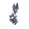
| Entry | Database: PDB / ID: 9cx9 | |||||||||||||||||||||||||||||||||||||||||||||||||||||||||||||||||||||
|---|---|---|---|---|---|---|---|---|---|---|---|---|---|---|---|---|---|---|---|---|---|---|---|---|---|---|---|---|---|---|---|---|---|---|---|---|---|---|---|---|---|---|---|---|---|---|---|---|---|---|---|---|---|---|---|---|---|---|---|---|---|---|---|---|---|---|---|---|---|---|
| Title | Structure of SH3 domain of Src in complex with beta-arrestin 1 | |||||||||||||||||||||||||||||||||||||||||||||||||||||||||||||||||||||
 Components Components |
| |||||||||||||||||||||||||||||||||||||||||||||||||||||||||||||||||||||
 Keywords Keywords | SIGNALING PROTEIN / GPCR signaling / arrestin / Src / SH3 | |||||||||||||||||||||||||||||||||||||||||||||||||||||||||||||||||||||
| Function / homology |  Function and homology information Function and homology informationV2 vasopressin receptor binding / alpha-1A adrenergic receptor binding / follicle-stimulating hormone receptor binding / TGFBR3 regulates TGF-beta signaling / G alpha (s) signalling events / sensory perception of touch / follicle-stimulating hormone signaling pathway / alpha-1B adrenergic receptor binding / protein phosphorylated amino acid binding / renal water retention ...V2 vasopressin receptor binding / alpha-1A adrenergic receptor binding / follicle-stimulating hormone receptor binding / TGFBR3 regulates TGF-beta signaling / G alpha (s) signalling events / sensory perception of touch / follicle-stimulating hormone signaling pathway / alpha-1B adrenergic receptor binding / protein phosphorylated amino acid binding / renal water retention / Defective AVP does not bind AVPR2 and causes neurohypophyseal diabetes insipidus (NDI) / Lysosome Vesicle Biogenesis / regulation of systemic arterial blood pressure by vasopressin / Vasopressin-like receptors / vasopressin receptor activity / Ub-specific processing proteases / renal water absorption / angiotensin receptor binding / AP-2 adaptor complex binding / MAP2K and MAPK activation / Golgi Associated Vesicle Biogenesis / Signaling by ERBB2 / Nuclear signaling by ERBB4 / Signaling by SCF-KIT / Regulation of KIT signaling / Signaling by EGFR / GAB1 signalosome / Regulation of gap junction activity / FCGR activation / PECAM1 interactions / Co-stimulation by CD28 / Co-inhibition by CTLA4 / EPHA-mediated growth cone collapse / Ephrin signaling / G alpha (i) signalling events / GP1b-IX-V activation signalling / Thrombin signalling through proteinase activated receptors (PARs) / VEGFR2 mediated cell proliferation / RET signaling / Receptor Mediated Mitophagy / : / ADP signalling through P2Y purinoceptor 1 / RAF activation / PIP3 activates AKT signaling / EPH-ephrin mediated repulsion of cells / PI5P, PP2A and IER3 Regulate PI3K/AKT Signaling / Downstream signal transduction / Downregulation of ERBB4 signaling / Cyclin D associated events in G1 / Regulation of RUNX3 expression and activity / Degradation of CDH1 / regulation of inositol trisphosphate biosynthetic process / MAP2K and MAPK activation / Integrin signaling / GRB2:SOS provides linkage to MAPK signaling for Integrins / : / MET activates PTK2 signaling / Extra-nuclear estrogen signaling / EPHB-mediated forward signaling / hemostasis / p130Cas linkage to MAPK signaling for integrins / VEGFA-VEGFR2 Pathway / Cargo recognition for clathrin-mediated endocytosis / Clathrin-mediated endocytosis / telencephalon development / clathrin-cargo adaptor activity / negative regulation of interleukin-8 production / connexin binding / desensitization of G protein-coupled receptor signaling pathway / regulation of G protein-coupled receptor signaling pathway / arrestin family protein binding / G protein-coupled receptor internalization / mitogen-activated protein kinase kinase binding / Thrombin signalling through proteinase activated receptors (PARs) / sensory perception / clathrin binding / stress fiber assembly / response to morphine / positive regulation of Rho protein signal transduction / negative regulation of intrinsic apoptotic signaling pathway / positive regulation of systemic arterial blood pressure / progesterone receptor signaling pathway / negative regulation of interleukin-6 production / pseudopodium / positive regulation of intracellular signal transduction / positive regulation of receptor internalization / negative regulation of Notch signaling pathway / phototransduction / positive regulation of vasoconstriction / endocytic vesicle / cysteine-type endopeptidase inhibitor activity / immune system process / activation of adenylate cyclase activity / cellular response to hormone stimulus / insulin-like growth factor receptor binding / clathrin-coated pit / response to cytokine / negative regulation of protein ubiquitination / nuclear estrogen receptor binding / positive regulation of insulin secretion involved in cellular response to glucose stimulus Similarity search - Function | |||||||||||||||||||||||||||||||||||||||||||||||||||||||||||||||||||||
| Biological species |    Homo sapiens (human) Homo sapiens (human) | |||||||||||||||||||||||||||||||||||||||||||||||||||||||||||||||||||||
| Method | ELECTRON MICROSCOPY / single particle reconstruction / cryo EM / Resolution: 3.34 Å | |||||||||||||||||||||||||||||||||||||||||||||||||||||||||||||||||||||
 Authors Authors | Pakharukova, N. / Bansia, H. / Lefkowitz, R.J. / des Georges, A. | |||||||||||||||||||||||||||||||||||||||||||||||||||||||||||||||||||||
| Funding support |  United States, European Union, United States, European Union,  France, 5items France, 5items
| |||||||||||||||||||||||||||||||||||||||||||||||||||||||||||||||||||||
 Citation Citation | Journal: Nat Commun / Year: 2026 Title: Mechanism of beta-arrestin 1 mediated Src activation via Src SH3 domain revealed by cryo-electron microscopy. Authors: Natalia Pakharukova / Brittany N Thomas / Harsh Bansia / Linus Li / Dana K Bassford / Rinat R Abzalimov / Jihee Kim / Alem W Kahsai / Biswaranjan Pani / Kunhong Xiao / Roni Ochakovski / ...Authors: Natalia Pakharukova / Brittany N Thomas / Harsh Bansia / Linus Li / Dana K Bassford / Rinat R Abzalimov / Jihee Kim / Alem W Kahsai / Biswaranjan Pani / Kunhong Xiao / Roni Ochakovski / Shibo Liu / Xingdong Zhang / Seungkirl Ahn / Amedee des Georges / Robert J Lefkowitz / Abstract: Beta-arrestins (βarrs) are key regulators and transducers of G-protein coupled receptor signaling; however, little is known of how βarrs communicate with their downstream effectors. Here, we ...Beta-arrestins (βarrs) are key regulators and transducers of G-protein coupled receptor signaling; however, little is known of how βarrs communicate with their downstream effectors. Here, we delineate structural mechanisms underlying βarr-mediated signal transduction. Using cryo-electron microscopy, we elucidate how βarr1 recruits and activates the non-receptor tyrosine kinase Src, a well-established signaling partner of βarrs. βarr1 engages Src SH3 through two distinct sites, each employing a different recognition mechanism: a polyproline motif in the N-domain and a non-proline-based interaction in the central crest region. At both sites βarr1 interacts with the aromatic surface of SH3, disrupting the autoinhibited conformation of Src and directly triggering its allosteric activation. This structural evidence establishes βarr1 as an active regulatory protein rather than a passive scaffold and suggests a potentially general mechanism for βarr-mediated signaling across diverse effectors. #1: Journal: bioRxiv / Year: 2025 Title: Mechanism of beta-arrestin 1 mediated Src activation via Src SH3 domain revealed by cryo-electron microscopy. Authors: Natalia Pakharukova / Brittany N Thomas / Harsh Bansia / Linus Li / Dana K Bassford / Rinat R Abzalimov / Jihee Kim / Alem W Kahsai / Biswaranjan Pani / Kunhong Xiao / Roni Ochakovski / ...Authors: Natalia Pakharukova / Brittany N Thomas / Harsh Bansia / Linus Li / Dana K Bassford / Rinat R Abzalimov / Jihee Kim / Alem W Kahsai / Biswaranjan Pani / Kunhong Xiao / Roni Ochakovski / Shibo Liu / Xingdong Zhang / Seungkirl Ahn / Amedee des Georges / Robert J Lefkowitz / Abstract: Beta-arrestins (βarrs) are key regulators and transducers of G-protein coupled receptor signaling; however, little is known of how βarrs communicate with their downstream effectors. Here, we report ...Beta-arrestins (βarrs) are key regulators and transducers of G-protein coupled receptor signaling; however, little is known of how βarrs communicate with their downstream effectors. Here, we report the first structural insights into the fundamental mechanisms driving βarr-mediated signal transduction. Using cryo-electron microscopy, we elucidate how βarr1 recruits and activates the non-receptor tyrosine kinase Src, the first identified signaling partner of βarrs. βarr1 engages Src SH3 through two distinct sites, each employing a different recognition mechanism: a polyproline motif in the N-domain and a non-proline-based interaction in the central crest region. At both sites βarr1 interacts with the aromatic surface of SH3, disrupting the autoinhibited conformation of Src and directly triggering its allosteric activation. This structural evidence establishes βarr1 as an active regulatory protein rather than a passive scaffold and suggests a potentially general mechanism for βarr-mediated signaling across diverse effectors. | |||||||||||||||||||||||||||||||||||||||||||||||||||||||||||||||||||||
| History |
|
- Structure visualization
| Structure viewer | Molecule: MolmilJmol/JSmol |
|---|
- Downloads & links
-Download
| PDBx/mmCIF format | 9cx9.cif.gz | 127.2 KB | Display | PDBx/mmCIF format |
|---|---|---|---|---|
| PDB format | pdb9cx9.ent.gz | 90.6 KB | Display | PDB format |
| PDBx/mmJSON format | 9cx9.json.gz | Tree view | PDBx/mmJSON format | |
| Others |  Other downloads Other downloads |
-Validation report
| Arichive directory | https://data.pdbj.org/pub/pdb/validation_reports/cx/9cx9ftp://data.pdbj.org/pub/pdb/validation_reports/cx/9cx9 | HTTPS FTP |
|---|
-Related structure data
| Related structure data |  45982MC  9bt8C  9cx3C C: citing same article ( M: map data used to model this data |
|---|---|
| Similar structure data |


-Links
 PDBj
PDBj


- Assembly
| Deposited unit | 
|
|---|---|
| 1 |
|
-Components
| #1: Antibody | Mass: 25512.354 Da / Num. of mol.: 1 Source method: isolated from a genetically manipulated source Source: (gene. exp.)  |
|---|---|
| #2: Antibody | Mass: 23435.064 Da / Num. of mol.: 1 Source method: isolated from a genetically manipulated source Source: (gene. exp.) |
| #3: Protein/peptide | Mass: 3550.936 Da / Num. of mol.: 1 / Fragment: UNP residues 343-371 / Source method: obtained synthetically Details: Synthetic phosphopeptide mimicking the C-tail of vasopressin 2 receptor Source: (synth.) Homo sapiens (human) / References: UniProt: P30518 |
| #4: Protein | Mass: 44049.160 Da / Num. of mol.: 1 Mutation: C59V, E92C, C125S, C140L, C150V, C242V, C251V, C269S Source method: isolated from a genetically manipulated source Source: (gene. exp.) Production host: References: UniProt: P29066 |
| #5: Protein | Mass: 9756.567 Da / Num. of mol.: 1 / Mutation: R95C Source method: isolated from a genetically manipulated source Source: (gene. exp.) Production host: References: UniProt: P00523, non-specific protein-tyrosine kinase |
| Has ligand of interest | N |
| Has protein modification | Y |
-Experimental details
-Experiment
| Experiment | Method: ELECTRON MICROSCOPY |
|---|---|
| EM experiment | Aggregation state: PARTICLE / 3D reconstruction method: single particle reconstruction |
- Sample preparation
| Component | Name: Beta-arrestin 1 bound to a G protein-coupled receptor phosphopeptide and antibody fragment Fab30 in complex with SH3 domain of Src Type: COMPLEX / Entity ID: all / Source: RECOMBINANT |
|---|---|
| Molecular weight | Experimental value: NO |
| Source (natural) | Organism: |
| Source (recombinant) | Organism: |
| Buffer solution | pH: 7.5 |
| Specimen | Conc.: 8 mg/ml / Embedding applied: NO / Shadowing applied: NO / Staining applied: NO / Vitrification applied: YES |
| Vitrification | Instrument: FEI VITROBOT MARK IV / Cryogen name: ETHANE / Humidity: 100 % / Chamber temperature: 277 K |
- Electron microscopy imaging
| Experimental equipment |  Model: Titan Krios / Image courtesy: FEI Company |
|---|---|
| Microscopy | Model: TFS KRIOS |
| Electron gun | Electron source:  FIELD EMISSION GUN / Accelerating voltage: 300 kV / Illumination mode: FLOOD BEAM FIELD EMISSION GUN / Accelerating voltage: 300 kV / Illumination mode: FLOOD BEAM |
| Electron lens | Mode: BRIGHT FIELD / Nominal defocus max: 2400 nm / Nominal defocus min: 800 nm |
| Image recording | Electron dose: 53.8 e/Å2 / Film or detector model: GATAN K3 (6k x 4k) |
- Processing
| EM software |
| ||||||||||||||||||||||||||||||||||||
|---|---|---|---|---|---|---|---|---|---|---|---|---|---|---|---|---|---|---|---|---|---|---|---|---|---|---|---|---|---|---|---|---|---|---|---|---|---|
| CTF correction | Type: PHASE FLIPPING AND AMPLITUDE CORRECTION | ||||||||||||||||||||||||||||||||||||
| 3D reconstruction | Resolution: 3.34 Å / Resolution method: FSC 0.143 CUT-OFF / Num. of particles: 345529 / Symmetry type: POINT | ||||||||||||||||||||||||||||||||||||
| Atomic model building | 3D fitting-ID: 1 / Source name: PDB / Type: experimental model
| ||||||||||||||||||||||||||||||||||||
| Refine LS restraints |
|