Activation and oligomerization of BAK protein / B cell negative selection / BAK complex / BH domain binding / negative regulation of endoplasmic reticulum calcium ion concentration / response to fungus / response to mycotoxin / Release of apoptotic factors from the mitochondria / apoptotic process involved in blood vessel morphogenesis / endocrine pancreas development ...Activation and oligomerization of BAK protein / B cell negative selection / BAK complex / BH domain binding / negative regulation of endoplasmic reticulum calcium ion concentration / response to fungus / response to mycotoxin / Release of apoptotic factors from the mitochondria / apoptotic process involved in blood vessel morphogenesis / endocrine pancreas development / post-embryonic camera-type eye morphogenesis / limb morphogenesis / establishment or maintenance of transmembrane electrochemical gradient / B cell apoptotic process / positive regulation of mitochondrial outer membrane permeabilization involved in apoptotic signaling pathway / negative regulation of mitochondrial outer membrane permeabilization involved in apoptotic signaling pathway / endoplasmic reticulum calcium ion homeostasis / calcium ion transport into cytosol / fibroblast apoptotic process / regulation of mitochondrial membrane permeability / mitochondrial fusion / myeloid cell homeostasis / response to UV-C / Bcl-2 family protein complex / thymocyte apoptotic process / porin activity / negative regulation of release of cytochrome c from mitochondria / positive regulation of IRE1-mediated unfolded protein response / pore complex / vagina development / B cell homeostasis / positive regulation of release of cytochrome c from mitochondria / positive regulation of proteolysis / intrinsic apoptotic signaling pathway in response to endoplasmic reticulum stress / animal organ regeneration / cellular response to unfolded protein / positive regulation of calcium ion transport into cytosol / Pyroptosis / blood vessel remodeling / extrinsic apoptotic signaling pathway in absence of ligand / heat shock protein binding / epithelial cell proliferation / intrinsic apoptotic signaling pathway / release of cytochrome c from mitochondria / response to gamma radiation / regulation of mitochondrial membrane potential / response to hydrogen peroxide / apoptotic signaling pathway / cellular response to mechanical stimulus / positive regulation of protein-containing complex assembly / intrinsic apoptotic signaling pathway in response to DNA damage / cellular response to UV / protein-folding chaperone binding / channel activity / response to ethanol / transmembrane transporter binding / mitochondrial outer membrane / regulation of cell cycle / positive regulation of apoptotic process / response to xenobiotic stimulus / protein heterodimerization activity / negative regulation of cell population proliferation / negative regulation of gene expression / apoptotic process / protein-containing complex binding / endoplasmic reticulum / protein homodimerization activity / mitochondrion / metal ion binding / identical protein binding / cytosol Similarity search - Function
Apoptosis regulator, Bcl-2, BH3 motif, conserved site / Apoptosis regulator, Bcl-2 family BH3 motif signature. / Apoptosis regulator, Bcl-2, BH1 motif, conserved site / Apoptosis regulator, Bcl-2 family BH1 motif signature. / Apoptosis regulator, Bcl-2, BH2 motif, conserved site / Apoptosis regulator, Bcl-2 family BH2 motif signature. / Bcl-2 family / BCL (B-Cell lymphoma); contains BH1, BH2 regions / Bcl2-like / Bcl-2, Bcl-2 homology region 1-3 ...Apoptosis regulator, Bcl-2, BH3 motif, conserved site / Apoptosis regulator, Bcl-2 family BH3 motif signature. / Apoptosis regulator, Bcl-2, BH1 motif, conserved site / Apoptosis regulator, Bcl-2 family BH1 motif signature. / Apoptosis regulator, Bcl-2, BH2 motif, conserved site / Apoptosis regulator, Bcl-2 family BH2 motif signature. / Bcl-2 family / BCL (B-Cell lymphoma); contains BH1, BH2 regions / Bcl2-like / Bcl-2, Bcl-2 homology region 1-3 / Apoptosis regulator proteins, Bcl-2 family / BCL2-like apoptosis inhibitors family profile. / Bcl-2-like superfamily Similarity search - Domain/homology
National Institutes of Health/National Institute of General Medical Sciences (NIH/NIGMS)
R01GM129470
United States
Citation
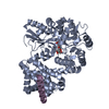
Journal: Mol Cell / Year: 2025 Title: Structural basis of BAK sequestration by MCL-1 in apoptosis. Authors: Shagun Srivastava / Giridhar Sekar / Adedolapo Ojoawo / Anup Aggarwal / Elisabeth Ferreira / Emiko Uchikawa / Meek Yang / Christy R Grace / Raja Dey / Yi-Lun Lin / Cristina D Guibao / ...Authors: Shagun Srivastava / Giridhar Sekar / Adedolapo Ojoawo / Anup Aggarwal / Elisabeth Ferreira / Emiko Uchikawa / Meek Yang / Christy R Grace / Raja Dey / Yi-Lun Lin / Cristina D Guibao / Seetharaman Jayaraman / Somnath Mukherjee / Anthony A Kossiakoff / Bin Dong / Alexander Myasnikov / Tudor Moldoveanu / Abstract: Apoptosis controls cell fate, ensuring tissue homeostasis and promoting disease when dysregulated. The rate-limiting step in apoptosis is mitochondrial poration by the effector B cell lymphoma 2 (BCL- ...Apoptosis controls cell fate, ensuring tissue homeostasis and promoting disease when dysregulated. The rate-limiting step in apoptosis is mitochondrial poration by the effector B cell lymphoma 2 (BCL-2) family proteins BAK and BAX, which are activated by initiator BCL-2 homology 3 (BH3)-only proteins (e.g., BIM) and inhibited by guardian BCL-2 family proteins (e.g., MCL-1). We integrated structural, biochemical, and pharmacological approaches to characterize the human prosurvival MCL-1:BAK complex assembled from their BCL-2 globular core domains. We reveal a canonical interaction with BAK BH3 bound to the hydrophobic groove of MCL-1 and disordered and highly dynamic BAK regions outside the complex interface. We predict similar conformations of activated effectors in complex with other guardians or effectors. The MCL-1:BAK complex is a major cancer drug target. We show that MCL-1 inhibitors are inefficient in neutralizing the MCL-1:BAK complex, requiring high doses to initiate apoptosis. Our study underscores the need to design superior clinical candidate MCL-1 inhibitors.
In the structure databanks used in Yorodumi, some data are registered as the other names, "COVID-19 virus" and "2019-nCoV". Here are the details of the virus and the list of structure data.
Jan 31, 2019. EMDB accession codes are about to change! (news from PDBe EMDB page)
EMDB accession codes are about to change! (news from PDBe EMDB page)
The allocation of 4 digits for EMDB accession codes will soon come to an end. Whilst these codes will remain in use, new EMDB accession codes will include an additional digit and will expand incrementally as the available range of codes is exhausted. The current 4-digit format prefixed with “EMD-” (i.e. EMD-XXXX) will advance to a 5-digit format (i.e. EMD-XXXXX), and so on. It is currently estimated that the 4-digit codes will be depleted around Spring 2019, at which point the 5-digit format will come into force.
The EM Navigator/Yorodumi systems omit the EMD- prefix.
Related info.:Q: What is EMD? / ID/Accession-code notation in Yorodumi/EM Navigator
Yorodumi is a browser for structure data from EMDB, PDB, SASBDB, etc.
This page is also the successor to EM Navigator detail page, and also detail information page/front-end page for Omokage search.
The word "yorodu" (or yorozu) is an old Japanese word meaning "ten thousand". "mi" (miru) is to see.
Related info.:EMDB / PDB / SASBDB / Comparison of 3 databanks / Yorodumi Search / Aug 31, 2016. New EM Navigator & Yorodumi / Yorodumi Papers / Jmol/JSmol / Function and homology information / Changes in new EM Navigator and Yorodumi
 Movie
Movie Controller

 Yorodumi
 Open data
 Basic information
 Components
 Keywords
 Function and homology information
 Homo sapiens (human)
 X-RAY DIFFRACTION /
  Authors
 United States, 1items
  Citation
 Structure visualization
 Downloads & links
 Other downloads

 PDBj
PDBj


 Assembly

 Mass: 18.015 Da / Num. of mol.: 161 / Source method: isolated from a natural source / Formula: H2O
 Sample preparation
 Processing