 Movie
Movie Controller

[English] 日本語
 Yorodumi
- PDB-9chv: cryo-EM structure of calcineurin-fused beta2 adrenergic receptor ... -
+ Open data

- Basic information
| Entry | Database: PDB / ID: 9chv | ||||||
|---|---|---|---|---|---|---|---|
| Title | cryo-EM structure of calcineurin-fused beta2 adrenergic receptor in apo state | ||||||
 Components Components |
| ||||||
 Keywords Keywords | MEMBRANE PROTEIN/HYDROLASE/ISOMERASE / GPCR / cryo-EM / calcineurin fusion / inactive state / MEMBRANE PROTEIN / MEMBRANE PROTEIN-HYDROLASE-ISOMERASE complex | ||||||
| Function / homology |  Function and homology information Function and homology informationpositive regulation of angiotensin-activated signaling pathway / negative regulation of angiotensin-activated signaling pathway / calcium-dependent protein serine/threonine phosphatase regulator activity / regulation of cell proliferation involved in kidney morphogenesis / CLEC7A (Dectin-1) induces NFAT activation / positive regulation of glomerulus development / negative regulation of calcium ion import across plasma membrane / negative regulation of signaling / calcium-dependent protein serine/threonine phosphatase activity / protein serine/threonine phosphatase complex ...positive regulation of angiotensin-activated signaling pathway / negative regulation of angiotensin-activated signaling pathway / calcium-dependent protein serine/threonine phosphatase regulator activity / regulation of cell proliferation involved in kidney morphogenesis / CLEC7A (Dectin-1) induces NFAT activation / positive regulation of glomerulus development / negative regulation of calcium ion import across plasma membrane / negative regulation of signaling / calcium-dependent protein serine/threonine phosphatase activity / protein serine/threonine phosphatase complex / positive regulation of saliva secretion / Calcineurin activates NFAT / calmodulin-dependent protein phosphatase activity / calcineurin complex / slit diaphragm / positive regulation of connective tissue replacement / positive regulation of calcium ion-dependent exocytosis of neurotransmitter / positive regulation of calcium ion import across plasma membrane / Ca2+ pathway / FCERI mediated Ca+2 mobilization / positive regulation of cardiac muscle hypertrophy in response to stress / macrolide binding / lung epithelial cell differentiation / negative regulation of dendrite morphogenesis / renal filtration / activin receptor binding / renal sodium ion absorption / regulation of skeletal muscle contraction by regulation of release of sequestered calcium ion / calcineurin-NFAT signaling cascade / cytoplasmic side of membrane / transforming growth factor beta receptor binding / TGFBR1 LBD Mutants in Cancer / positive regulation of calcineurin-NFAT signaling cascade / type I transforming growth factor beta receptor binding / negative regulation of activin receptor signaling pathway / myelination in peripheral nervous system / transition between fast and slow fiber / signaling receptor inhibitor activity / heart trabecula formation / positive regulation of osteoclast differentiation / beta2-adrenergic receptor activity / cardiac muscle hypertrophy in response to stress / I-SMAD binding / positive regulation of mini excitatory postsynaptic potential / AMPA selective glutamate receptor signaling pathway / norepinephrine-epinephrine-mediated vasodilation involved in regulation of systemic arterial blood pressure / adenylate cyclase-inhibiting adrenergic receptor signaling pathway / positive regulation of autophagosome maturation / heat generation / norepinephrine binding / regulation of amyloid precursor protein catabolic process / Adrenoceptors / terminal cisterna / ryanodine receptor complex / negative regulation of smooth muscle contraction / regulation of synaptic vesicle cycle / positive regulation of cardiac muscle cell contraction / positive regulation of activated T cell proliferation / positive regulation of lipophagy / protein dephosphorylation / negative regulation of multicellular organism growth / negative regulation of G protein-coupled receptor signaling pathway / 'de novo' protein folding / branching involved in blood vessel morphogenesis / adrenergic receptor signaling pathway / ventricular cardiac muscle tissue morphogenesis / regulation of postsynaptic neurotransmitter receptor internalization / protein-serine/threonine phosphatase / FK506 binding / CLEC7A (Dectin-1) induces NFAT activation / diet induced thermogenesis / response to psychosocial stress / endosome to lysosome transport / positive regulation of cardiac muscle hypertrophy / protein serine/threonine phosphatase activity / TGF-beta receptor signaling activates SMADs / parallel fiber to Purkinje cell synapse / calcineurin-mediated signaling / positive regulation of cAMP/PKA signal transduction / smooth muscle contraction / negative regulation of cardiac muscle cell apoptotic process / adenylate cyclase binding / mTORC1-mediated signalling / epithelial to mesenchymal transition / Calcineurin activates NFAT / epidermis development / Activation of BAD and translocation to mitochondria / DARPP-32 events / positive regulation of osteoblast differentiation / potassium channel regulator activity / regulation of immune response / positive regulation of endocytosis / bone resorption / positive regulation of bone mineralization / multicellular organismal response to stress / postsynaptic modulation of chemical synaptic transmission / neuronal dense core vesicle / phosphatase binding / heart morphogenesis / keratinocyte differentiation Similarity search - Function | ||||||
| Biological species |  Homo sapiens (human) Homo sapiens (human) | ||||||
| Method | ELECTRON MICROSCOPY / single particle reconstruction / cryo EM / Resolution: 3.95 Å | ||||||
 Authors Authors | Xu, J. / Chen, G. / Du, Y. / Kobilka, B.K. | ||||||
| Funding support | 1items
| ||||||
 Citation Citation | Journal: Proc Natl Acad Sci U S A / Year: 2024 Title: Calcineurin-fusion facilitates cryo-EM structure determination of a Family A GPCR. Authors: Jun Xu / Geng Chen / Haoqing Wang / Sheng Cao / Jie Heng / Xavier Deupi / Yang Du / Brian K Kobilka /    Abstract: Advances in singe-particle cryo-electron microscopy (cryo-EM) have made it possible to solve the structures of numerous Family A and Family B G protein-coupled receptors (GPCRs) in complex with G ...Advances in singe-particle cryo-electron microscopy (cryo-EM) have made it possible to solve the structures of numerous Family A and Family B G protein-coupled receptors (GPCRs) in complex with G proteins and arrestins, as well as several Family C GPCRs. Determination of these structures has been facilitated by the presence of large extramembrane components (such as G protein, arrestin, or Venus flytrap domains) in these complexes that aid in particle alignment during the processing of the cryo-EM data. In contrast, determination of the inactive state structure of Family A GPCRs is more challenging due to the relatively small size of the seven transmembrane domain (7TM) and to the surrounding detergent micelle that, in the absence of other features, make particle alignment impossible. Here, we describe an alternative protein engineering strategy where the heterodimeric protein calcineurin is fused to a GPCR by three points of attachment, the cytoplasmic ends of TM5, TM6, and TM7. This three-point attachment provides a more rigid link with the GPCR transmembrane domain that facilitates particle alignment during data processing, allowing us to determine the structures of the β adrenergic receptor (βAR) in the apo, antagonist-bound, and agonist-bound states. We expect that this fusion strategy may have broad application in cryo-EM structural determination of other Family A GPCRs. | ||||||
| History |
|
- Structure visualization
| Structure viewer | Molecule: MolmilJmol/JSmol |
|---|
- Downloads & links
-Download
| PDBx/mmCIF format | 9chv.cif.gz | 167.3 KB | Display | PDBx/mmCIF format |
|---|---|---|---|---|
| PDB format | pdb9chv.ent.gz | 124.9 KB | Display | PDB format |
| PDBx/mmJSON format | 9chv.json.gz | Tree view | PDBx/mmJSON format | |
| Others |  Other downloads Other downloads |
-Validation report
| Arichive directory | https://data.pdbj.org/pub/pdb/validation_reports/ch/9chvftp://data.pdbj.org/pub/pdb/validation_reports/ch/9chv | HTTPS FTP |
|---|
-Related structure data
| Related structure data |  45603MC  9chuC  9chxC C: citing same article ( M: map data used to model this data |
|---|---|
| Similar structure data |


-Links
 PDBj
PDBj


- Assembly
| Deposited unit | 
|
|---|---|
| 1 |
|
-Components
| #1: Protein | Mass: 51309.711 Da / Num. of mol.: 1 Source method: isolated from a genetically manipulated source Details: residues 29-354 (Uniprot numbering) of the beta2-AR with the third cytoplasmic domain replaced with residues 16-170 (Uniprot numbering) of calcineurin subunit B Source: (gene. exp.) Homo sapiens (human) / Gene: ADRB2, ADRB2R, B2AR, PPP3R1, CNA2, CNB / Production host:   Spodoptera frugiperda (fall armyworm) / References: UniProt: P07550, UniProt: P63098 Spodoptera frugiperda (fall armyworm) / References: UniProt: P07550, UniProt: P63098 |
|---|---|
| #2: Protein | Mass: 42627.785 Da / Num. of mol.: 1 / Fragment: residues 1-370 Source method: isolated from a genetically manipulated source Source: (gene. exp.) Spodoptera frugiperda (fall armyworm)References: UniProt: P63328, protein-serine/threonine phosphatase |
| #3: Protein | Mass: 11967.705 Da / Num. of mol.: 1 Source method: isolated from a genetically manipulated source Source: (gene. exp.) Homo sapiens (human) / Gene: FKBP1A, FKBP1, FKBP12Production host:  References: UniProt: P62942, peptidylprolyl isomerase |
| #4: Chemical | ChemComp-FK5 / 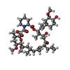  Mass: 804.018 Da / Num. of mol.: 1 / Source method: obtained synthetically / Formula: C44H69NO12 / Feature type: SUBJECT OF INVESTIGATION / Comment: medication*YM Mass: 804.018 Da / Num. of mol.: 1 / Source method: obtained synthetically / Formula: C44H69NO12 / Feature type: SUBJECT OF INVESTIGATION / Comment: medication*YM |
| Has ligand of interest | Y |
| Has protein modification | Y |
-Experimental details
-Experiment
| Experiment | Method: ELECTRON MICROSCOPY |
|---|---|
| EM experiment | Aggregation state: PARTICLE / 3D reconstruction method: single particle reconstruction |
- Sample preparation
| Component |
| ||||||||||||||||||||||||||||||
|---|---|---|---|---|---|---|---|---|---|---|---|---|---|---|---|---|---|---|---|---|---|---|---|---|---|---|---|---|---|---|---|
| Source (natural) |
| ||||||||||||||||||||||||||||||
| Source (recombinant) |
| ||||||||||||||||||||||||||||||
| Buffer solution | pH: 7.5 | ||||||||||||||||||||||||||||||
| Specimen | Embedding applied: NO / Shadowing applied: NO / Staining applied: NO / Vitrification applied: YES | ||||||||||||||||||||||||||||||
| Vitrification | Cryogen name: ETHANE |
- Electron microscopy imaging
| Experimental equipment |  Model: Titan Krios / Image courtesy: FEI Company |
|---|---|
| Microscopy | Model: TFS KRIOS |
| Electron gun | Electron source:  FIELD EMISSION GUN / Accelerating voltage: 300 kV / Illumination mode: OTHER FIELD EMISSION GUN / Accelerating voltage: 300 kV / Illumination mode: OTHER |
| Electron lens | Mode: BRIGHT FIELD / Nominal defocus max: 2000 nm / Nominal defocus min: 1000 nm |
| Image recording | Electron dose: 48 e/Å2 / Film or detector model: GATAN K2 QUANTUM (4k x 4k) |
- Processing
| EM software | Name: PHENIX / Version: 1.18_3861: / Category: model refinement | ||||||||||||||||||||||||
|---|---|---|---|---|---|---|---|---|---|---|---|---|---|---|---|---|---|---|---|---|---|---|---|---|---|
| CTF correction | Type: PHASE FLIPPING AND AMPLITUDE CORRECTION | ||||||||||||||||||||||||
| 3D reconstruction | Resolution: 3.95 Å / Resolution method: FSC 0.143 CUT-OFF / Num. of particles: 279590 / Symmetry type: POINT | ||||||||||||||||||||||||
| Refine LS restraints |
|
