Movie
Movie Controller

[English] 日本語
 Yorodumi
- EMDB-45604: cryo-EM structure of calcineurin-fused beta2 adrenergic receptor ... -
+ Open data

- Basic information
| Entry |  | |||||||||
|---|---|---|---|---|---|---|---|---|---|---|
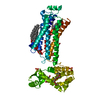
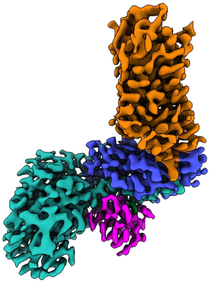
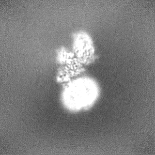
| Title | cryo-EM structure of calcineurin-fused beta2 adrenergic receptor in carazolol bound inactive state | |||||||||
 Map data Map data | ||||||||||
 Sample Sample |
| |||||||||
 Keywords Keywords | GPCR / cryo-EM / calcineurin fusion / inactive state / MEMBRANE PROTEIN / MEMBRANE PROTEIN-HYDROLASE-ISOMERASE complex | |||||||||
| Function / homology |  Function and homology information Function and homology informationpositive regulation of angiotensin-activated signaling pathway / negative regulation of angiotensin-activated signaling pathway / calcium-dependent protein serine/threonine phosphatase regulator activity / regulation of cell proliferation involved in kidney morphogenesis / CLEC7A (Dectin-1) induces NFAT activation / positive regulation of glomerulus development / negative regulation of calcium ion import across plasma membrane / negative regulation of signaling / calcium-dependent protein serine/threonine phosphatase activity / protein serine/threonine phosphatase complex ...positive regulation of angiotensin-activated signaling pathway / negative regulation of angiotensin-activated signaling pathway / calcium-dependent protein serine/threonine phosphatase regulator activity / regulation of cell proliferation involved in kidney morphogenesis / CLEC7A (Dectin-1) induces NFAT activation / positive regulation of glomerulus development / negative regulation of calcium ion import across plasma membrane / negative regulation of signaling / calcium-dependent protein serine/threonine phosphatase activity / protein serine/threonine phosphatase complex / positive regulation of saliva secretion / Calcineurin activates NFAT / calmodulin-dependent protein phosphatase activity / calcineurin complex / slit diaphragm / positive regulation of connective tissue replacement / positive regulation of calcium ion-dependent exocytosis of neurotransmitter / positive regulation of calcium ion import across plasma membrane / Ca2+ pathway / FCERI mediated Ca+2 mobilization / positive regulation of cardiac muscle hypertrophy in response to stress / macrolide binding / lung epithelial cell differentiation / negative regulation of dendrite morphogenesis / renal filtration / activin receptor binding / renal sodium ion absorption / regulation of skeletal muscle contraction by regulation of release of sequestered calcium ion / calcineurin-NFAT signaling cascade / cytoplasmic side of membrane / transforming growth factor beta receptor binding / TGFBR1 LBD Mutants in Cancer / positive regulation of calcineurin-NFAT signaling cascade / type I transforming growth factor beta receptor binding / negative regulation of activin receptor signaling pathway / myelination in peripheral nervous system / transition between fast and slow fiber / signaling receptor inhibitor activity / heart trabecula formation / positive regulation of osteoclast differentiation / beta2-adrenergic receptor activity / cardiac muscle hypertrophy in response to stress / I-SMAD binding / positive regulation of mini excitatory postsynaptic potential / AMPA selective glutamate receptor signaling pathway / norepinephrine-epinephrine-mediated vasodilation involved in regulation of systemic arterial blood pressure / adenylate cyclase-inhibiting adrenergic receptor signaling pathway / positive regulation of autophagosome maturation / heat generation / norepinephrine binding / regulation of amyloid precursor protein catabolic process / Adrenoceptors / terminal cisterna / ryanodine receptor complex / negative regulation of smooth muscle contraction / regulation of synaptic vesicle cycle / positive regulation of cardiac muscle cell contraction / positive regulation of activated T cell proliferation / positive regulation of lipophagy / protein dephosphorylation / negative regulation of multicellular organism growth / negative regulation of G protein-coupled receptor signaling pathway / 'de novo' protein folding / branching involved in blood vessel morphogenesis / adrenergic receptor signaling pathway / ventricular cardiac muscle tissue morphogenesis / regulation of postsynaptic neurotransmitter receptor internalization / protein-serine/threonine phosphatase / FK506 binding / CLEC7A (Dectin-1) induces NFAT activation / diet induced thermogenesis / response to psychosocial stress / endosome to lysosome transport / positive regulation of cardiac muscle hypertrophy / protein serine/threonine phosphatase activity / TGF-beta receptor signaling activates SMADs / parallel fiber to Purkinje cell synapse / calcineurin-mediated signaling / positive regulation of cAMP/PKA signal transduction / smooth muscle contraction / negative regulation of cardiac muscle cell apoptotic process / adenylate cyclase binding / mTORC1-mediated signalling / epithelial to mesenchymal transition / Calcineurin activates NFAT / epidermis development / Activation of BAD and translocation to mitochondria / DARPP-32 events / positive regulation of osteoblast differentiation / potassium channel regulator activity / regulation of immune response / positive regulation of endocytosis / bone resorption / positive regulation of bone mineralization / multicellular organismal response to stress / postsynaptic modulation of chemical synaptic transmission / neuronal dense core vesicle / phosphatase binding / heart morphogenesis / keratinocyte differentiation Similarity search - Function | |||||||||
| Biological species |  Homo sapiens (human) / Homo sapiens (human) /  | |||||||||
| Method | single particle reconstruction / cryo EM / Resolution: 3.5 Å | |||||||||
 Authors Authors | Xu J / Chen G / Du Y / Kobilka BK | |||||||||
| Funding support | 1 items
| |||||||||
 Citation Citation | Journal: Proc Natl Acad Sci U S A / Year: 2024 Title: Calcineurin-fusion facilitates cryo-EM structure determination of a Family A GPCR. Authors: Jun Xu / Geng Chen / Haoqing Wang / Sheng Cao / Jie Heng / Xavier Deupi / Yang Du / Brian K Kobilka /    Abstract: Advances in singe-particle cryo-electron microscopy (cryo-EM) have made it possible to solve the structures of numerous Family A and Family B G protein-coupled receptors (GPCRs) in complex with G ...Advances in singe-particle cryo-electron microscopy (cryo-EM) have made it possible to solve the structures of numerous Family A and Family B G protein-coupled receptors (GPCRs) in complex with G proteins and arrestins, as well as several Family C GPCRs. Determination of these structures has been facilitated by the presence of large extramembrane components (such as G protein, arrestin, or Venus flytrap domains) in these complexes that aid in particle alignment during the processing of the cryo-EM data. In contrast, determination of the inactive state structure of Family A GPCRs is more challenging due to the relatively small size of the seven transmembrane domain (7TM) and to the surrounding detergent micelle that, in the absence of other features, make particle alignment impossible. Here, we describe an alternative protein engineering strategy where the heterodimeric protein calcineurin is fused to a GPCR by three points of attachment, the cytoplasmic ends of TM5, TM6, and TM7. This three-point attachment provides a more rigid link with the GPCR transmembrane domain that facilitates particle alignment during data processing, allowing us to determine the structures of the β adrenergic receptor (βAR) in the apo, antagonist-bound, and agonist-bound states. We expect that this fusion strategy may have broad application in cryo-EM structural determination of other Family A GPCRs. | |||||||||
| History |
|
- Structure visualization
| Supplemental images |
|---|
- Downloads & links
-EMDB archive
| Map data | emd_45604.map.gz | 117.8 MB | EMDB map data format | |
|---|---|---|---|---|
| Header (meta data) | emd-45604-v30.xmlemd-45604.xml | 18.8 KB 18.8 KB | Display Display | EMDB header |
| Images |  emd_45604.png emd_45604.png | 130.1 KB | ||
| Filedesc metadata | emd-45604.cif.gz | 6.5 KB | ||
| Others | emd_45604_half_map_1.map.gzemd_45604_half_map_2.map.gz | 115.9 MB 115.9 MB | ||
| Archive directory |  http://ftp.pdbj.org/pub/emdb/structures/EMD-45604ftp://ftp.pdbj.org/pub/emdb/structures/EMD-45604 http://ftp.pdbj.org/pub/emdb/structures/EMD-45604ftp://ftp.pdbj.org/pub/emdb/structures/EMD-45604 | HTTPS FTP |
-Related structure data
| Related structure data |  9chxMC  9chuC  9chvC M: atomic model generated by this map C: citing same article ( |
|---|---|
| Similar structure data |


-Links
| EMDB pages | EMDB (EBI/PDBe) / EMDataResource |
|---|---|
| Related items in Molecule of the Month |


-Map
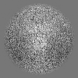
| File | Download / File: emd_45604.map.gz / Format: CCP4 / Size: 125 MB / Type: IMAGE STORED AS FLOATING POINT NUMBER (4 BYTES) | ||||||||||||||||||||||||||||||||||||
|---|---|---|---|---|---|---|---|---|---|---|---|---|---|---|---|---|---|---|---|---|---|---|---|---|---|---|---|---|---|---|---|---|---|---|---|---|---|
| Projections & slices | Image control
Images are generated by Spider. | ||||||||||||||||||||||||||||||||||||
| Voxel size | X=Y=Z: 0.85 Å | ||||||||||||||||||||||||||||||||||||
| Density |
| ||||||||||||||||||||||||||||||||||||
| Symmetry | Space group: 1 | ||||||||||||||||||||||||||||||||||||
| Details | EMDB XML:
|
 Z (Sec.)
 Y (Row.)
 X (Col.)


-Supplemental data
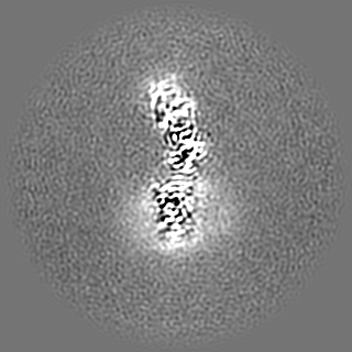
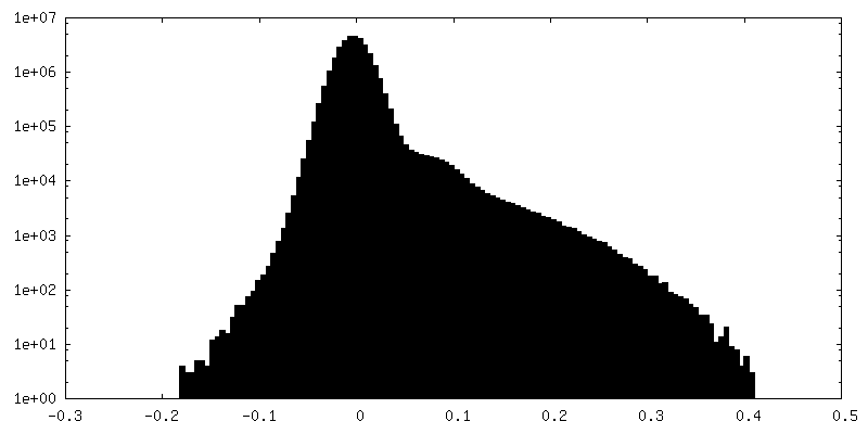
-Half map: #1
| File | emd_45604_half_map_1.map | ||||||||||||
|---|---|---|---|---|---|---|---|---|---|---|---|---|---|
| Projections & Slices |
| ||||||||||||
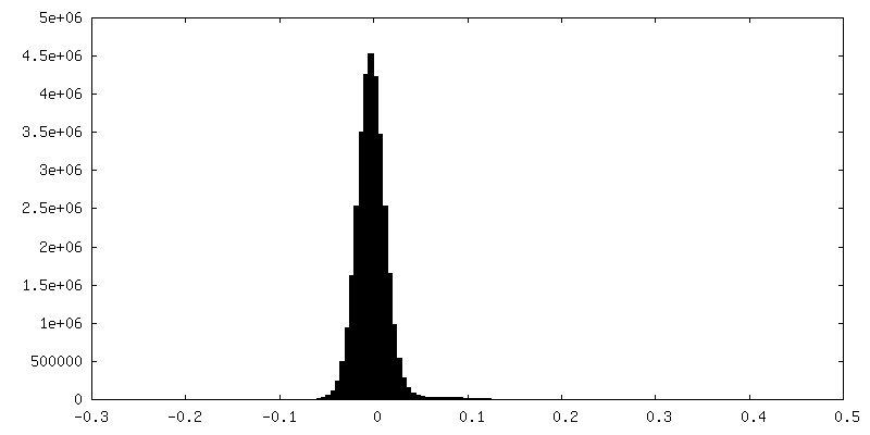
| Density Histograms |
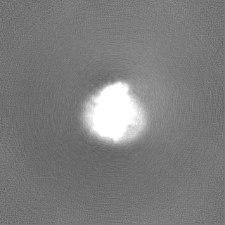


-Half map: #2
| File | emd_45604_half_map_2.map | ||||||||||||
|---|---|---|---|---|---|---|---|---|---|---|---|---|---|
| Projections & Slices |
| ||||||||||||
| Density Histograms |


- Sample components
-Entire : calcineurin fused beta2AR in complex with FKBP12
| Entire | Name: calcineurin fused beta2AR in complex with FKBP12 |
|---|---|
| Components |
|
-Supramolecule #1: calcineurin fused beta2AR in complex with FKBP12
| Supramolecule | Name: calcineurin fused beta2AR in complex with FKBP12 / type: complex / ID: 1 / Parent: 0 / Macromolecule list: #1-#3 |
|---|
-Supramolecule #2: calcineurin fused beta2AR
| Supramolecule | Name: calcineurin fused beta2AR / type: complex / ID: 2 / Parent: 1 / Macromolecule list: #1 |
|---|---|
| Source (natural) | Organism: Homo sapiens (human) |
-Supramolecule #3: Protein phosphatase 3 catalytic subunit alpha
| Supramolecule | Name: Protein phosphatase 3 catalytic subunit alpha / type: complex / ID: 3 / Parent: 1 / Macromolecule list: #2 |
|---|---|
| Source (natural) | Organism: |
-Supramolecule #4: FKBP1A
| Supramolecule | Name: FKBP1A / type: complex / ID: 4 / Parent: 1 / Macromolecule list: #3 |
|---|---|
| Source (natural) | Organism: Homo sapiens (human) |
-Macromolecule #1: Beta-2 adrenergic receptor,Calcineurin subunit B type 1
| Macromolecule | Name: Beta-2 adrenergic receptor,Calcineurin subunit B type 1 type: protein_or_peptide / ID: 1 Details: residues 29-354 (Uniprot numbering) of the beta2-AR with the third cytoplasmic domain replaced with residues 16-170 (Uniprot numbering) of calcineurin subunit B Number of copies: 1 / Enantiomer: LEVO |
|---|---|
| Source (natural) | Organism: Homo sapiens (human) |
| Molecular weight | Theoretical: 51.309711 KDa |
| Recombinant expression | Organism:   Spodoptera frugiperda (fall armyworm) Spodoptera frugiperda (fall armyworm) |
| Sequence | String: DEVWVVGMGI VMSLIVLAIV FGNVLVITAI AKFERLQTVT NYFITSLACA DLVMGLAVVP FGAAHILMKM WTFGNFWCEF WTSIDVLCV TASIETLCVI AVDRYFAITS PFKYQSLLTK NKARVIILMV WIVSGLTSFL PIQMHWYRAT HQEAINCYAE E TCCDFFTN ...String: DEVWVVGMGI VMSLIVLAIV FGNVLVITAI AKFERLQTVT NYFITSLACA DLVMGLAVVP FGAAHILMKM WTFGNFWCEF WTSIDVLCV TASIETLCVI AVDRYFAITS PFKYQSLLTK NKARVIILMV WIVSGLTSFL PIQMHWYRAT HQEAINCYAE E TCCDFFTN QAYAIASSIV SFYVPLVIMV FVYSRVFQEA KRQLDADEIK RLGKRFKKLD LDNSGSLSVE EFMSLPELQQ NP LVQRVID IFDTDGNGEV DFKEFIEGVS QFSVKGDKEQ KLRFAFRIYD MDKDGYISNG ELFQVLKMMV GNNLKDTQLQ QIV DKTIIN ADKDGDGRIS FEEFCAVVGG LDIHKKMVVD VKFCLKEHKA LKTLGIIMGT FTLCWLPFFI VNIVHVIQDN LIRK EVYIL LNWIGYVNSG FNPLIYCRSP DFRIAFQELL CLRRDDLKAY GNGY UniProtKB: Beta-2 adrenergic receptor, Calcineurin subunit B type 1, Beta-2 adrenergic receptor |
-Macromolecule #2: Protein phosphatase 3 catalytic subunit alpha
| Macromolecule | Name: Protein phosphatase 3 catalytic subunit alpha / type: protein_or_peptide / ID: 2 / Number of copies: 1 / Enantiomer: LEVO / EC number: protein-serine/threonine phosphatase |
|---|---|
| Source (natural) | Organism: |
| Molecular weight | Theoretical: 42.627785 KDa |
| Recombinant expression | Organism: Spodoptera frugiperda (fall armyworm) |
| Sequence | String: SEPKAIDPKL STTDRVVKAV PFPPSHRLTA KEVFDNDGKP RVDILKAHLM KEGRLEESVA LRIITEGASI LRQEKNLLDI DAPVTVCGD IHGQFFDLMK LFEVGGSPAN TRYLFLGDYV DRGYFSIECV LYLWALKILY PKTLFLLRGN HECRHLTEYF T FKQECKIK ...String: SEPKAIDPKL STTDRVVKAV PFPPSHRLTA KEVFDNDGKP RVDILKAHLM KEGRLEESVA LRIITEGASI LRQEKNLLDI DAPVTVCGD IHGQFFDLMK LFEVGGSPAN TRYLFLGDYV DRGYFSIECV LYLWALKILY PKTLFLLRGN HECRHLTEYF T FKQECKIK YSERVYDACM DAFDCLPLAA LMNQQFLCVH GGLSPEINTL DDIRKLDRFK EPPAYGPMCD ILWSDPLEDF GN EKTQEHF THNTVRGCSY FYSYPAVCDF LQHNNLLSIL RAHEAQDAGY RMYRKSQTTG FPSLITIFSA PNYLDVYNNK AAV LKYENN VMNIRQFNCS PHPYWLPNFM DVFTWSLPFV GEKVTEMLVN VLNI UniProtKB: Protein phosphatase 3 catalytic subunit alpha |
-Macromolecule #3: Peptidyl-prolyl cis-trans isomerase FKBP1A
| Macromolecule | Name: Peptidyl-prolyl cis-trans isomerase FKBP1A / type: protein_or_peptide / ID: 3 / Number of copies: 1 / Enantiomer: LEVO / EC number: peptidylprolyl isomerase |
|---|---|
| Source (natural) | Organism: Homo sapiens (human) |
| Molecular weight | Theoretical: 11.967705 KDa |
| Recombinant expression | Organism:  |
| Sequence | String: MGVQVETISP GDGRTFPKRG QTCVVHYTGM LEDGKKFDSS RDRNKPFKFM LGKQEVIRGW EEGVAQMSVG QRAKLTISPD YAYGATGHP GIIPPHATLV FDVELLKLE UniProtKB: Peptidyl-prolyl cis-trans isomerase FKBP1A |
-Macromolecule #4: (2S)-1-(9H-Carbazol-4-yloxy)-3-(isopropylamino)propan-2-ol
| Macromolecule | Name: (2S)-1-(9H-Carbazol-4-yloxy)-3-(isopropylamino)propan-2-ol type: ligand / ID: 4 / Number of copies: 1 / Formula: CAU |
|---|---|
| Molecular weight | Theoretical: 298.379 Da |
| Chemical component information |  ChemComp-CAU: |
-Macromolecule #5: 8-DEETHYL-8-[BUT-3-ENYL]-ASCOMYCIN
| Macromolecule | Name: 8-DEETHYL-8-[BUT-3-ENYL]-ASCOMYCIN / type: ligand / ID: 5 / Number of copies: 1 / Formula: FK5 |
|---|---|
| Molecular weight | Theoretical: 804.018 Da |
| Chemical component information |  ChemComp-FK5: |
-Experimental details
-Structure determination
| Method | cryo EM |
|---|---|
 Processing Processing | single particle reconstruction |
| Aggregation state | particle |
-Sample preparation
| Buffer | pH: 7.5 |
|---|---|
| Vitrification | Cryogen name: ETHANE |
- Electron microscopy
| Microscope | TFS KRIOS |
|---|---|
| Image recording | Film or detector model: GATAN K2 QUANTUM (4k x 4k) / Average electron dose: 48.0 e/Å2 |
| Electron beam | Acceleration voltage: 300 kV / Electron source:  FIELD EMISSION GUN FIELD EMISSION GUN |
| Electron optics | Illumination mode: OTHER / Imaging mode: BRIGHT FIELD / Nominal defocus max: 2.0 µm / Nominal defocus min: 1.0 µm |
| Experimental equipment |  Model: Titan Krios / Image courtesy: FEI Company |
-Image processing
| Startup model | Type of model: PDB ENTRY PDB model - PDB ID: |
|---|---|
| Final reconstruction | Resolution.type: BY AUTHOR / Resolution: 3.5 Å / Resolution method: FSC 0.143 CUT-OFF / Number images used: 276093 |
| Initial angle assignment | Type: MAXIMUM LIKELIHOOD |
| Final angle assignment | Type: NOT APPLICABLE |