 Movie
Movie Controller

[English] 日本語
 Yorodumi
- PDB-9cgd: Human DJ-1, 10 sec mixing with methylglyoxal, pink beam time-reso... -
+ Open data

- Basic information
| Entry | Database: PDB / ID: 9cgd | |||||||||
|---|---|---|---|---|---|---|---|---|---|---|
| Title | Human DJ-1, 10 sec mixing with methylglyoxal, pink beam time-resolved serial crystallography, CrystFEL processed | |||||||||
 Components Components | Parkinson disease protein 7 | |||||||||
 Keywords Keywords | HYDROLASE / Glutathione-independent glyoxalase / mix-and-inject serial crystallography / Laue diffraction | |||||||||
| Function / homology |  Function and homology information Function and homology informationpositive regulation of acute inflammatory response to antigenic stimulus / tyrosine 3-monooxygenase activator activity / cellular response to glyoxal / L-dopa decarboxylase activator activity / detoxification of hydrogen peroxide / : / detection of oxidative stress / : / guanine deglycation, glyoxal removal / cellular detoxification of methylglyoxal ...positive regulation of acute inflammatory response to antigenic stimulus / tyrosine 3-monooxygenase activator activity / cellular response to glyoxal / L-dopa decarboxylase activator activity / detoxification of hydrogen peroxide / : / detection of oxidative stress / : / guanine deglycation, glyoxal removal / cellular detoxification of methylglyoxal / regulation of supramolecular fiber organization / negative regulation of death-inducing signaling complex assembly / negative regulation of TRAIL-activated apoptotic signaling pathway / : / glyoxalase (glycolic acid-forming) activity / negative regulation of protein K48-linked deubiquitination / negative regulation of nitrosative stress-induced intrinsic apoptotic signaling pathway / glycolate biosynthetic process / glyoxal metabolic process / guanine deglycation / detoxification of mercury ion / ubiquitin-protein transferase inhibitor activity / protein deglycase / hydrogen peroxide metabolic process / mercury ion binding / methylglyoxal metabolic process / protein deglycase activity / positive regulation of autophagy of mitochondrion / superoxide dismutase copper chaperone activity / oxidoreductase activity, acting on peroxide as acceptor / positive regulation of dopamine biosynthetic process / positive regulation of mitochondrial electron transport, NADH to ubiquinone / lactate biosynthetic process / negative regulation of hydrogen peroxide-induced neuron intrinsic apoptotic signaling pathway / protein repair / peptidase inhibitor activity / peroxiredoxin activity / cellular detoxification of aldehyde / small protein activating enzyme binding / Hydrolases; Acting on ester bonds; Thioester hydrolases / regulation of oxidative stress-induced neuron intrinsic apoptotic signaling pathway / detoxification of copper ion / negative regulation of protein sumoylation / positive regulation of oxidative stress-induced intrinsic apoptotic signaling pathway / negative regulation of protein export from nucleus / cupric ion binding / regulation of androgen receptor signaling pathway / negative regulation of oxidative stress-induced neuron intrinsic apoptotic signaling pathway / membrane hyperpolarization / Hydrolases; Acting on carbon-nitrogen bonds, other than peptide bonds; In linear amides / oxygen sensor activity / insulin secretion / androgen receptor signaling pathway / nuclear androgen receptor binding / negative regulation of intrinsic apoptotic signaling pathway in response to hydrogen peroxide / ubiquitin-like protein conjugating enzyme binding / ubiquitin-specific protease binding / cytokine binding / positive regulation of reactive oxygen species biosynthetic process / dopamine uptake involved in synaptic transmission / cuprous ion binding / signaling receptor activator activity / regulation of synaptic vesicle endocytosis / single fertilization / membrane depolarization / negative regulation of endoplasmic reticulum stress-induced intrinsic apoptotic signaling pathway / negative regulation of oxidative stress-induced intrinsic apoptotic signaling pathway / negative regulation of reactive oxygen species biosynthetic process / removal of superoxide radicals / SUMOylation of transcription cofactors / negative regulation of protein ubiquitination / negative regulation of proteasomal ubiquitin-dependent protein catabolic process / regulation of neuron apoptotic process / negative regulation of extrinsic apoptotic signaling pathway / positive regulation of interleukin-8 production / regulation of mitochondrial membrane potential / adult locomotory behavior / adherens junction / mitochondrion organization / centriole / positive regulation of protein-containing complex assembly / Late endosomal microautophagy / mitochondrial intermembrane space / PML body / positive regulation of protein localization to nucleus / autophagy / positive regulation of reactive oxygen species metabolic process / enzyme activator activity / kinase binding / cellular response to hydrogen peroxide / Chaperone Mediated Autophagy / Aggrephagy / synaptic vesicle / peptidase activity / glucose homeostasis / cellular response to oxidative stress / cell body / regulation of inflammatory response / response to oxidative stress / scaffold protein binding Similarity search - Function | |||||||||
| Biological species |  Homo sapiens (human) Homo sapiens (human) | |||||||||
| Method |  X-RAY DIFFRACTION / SYNCHROTRON / FOURIER SYNTHESIS / Resolution: 1.97 Å X-RAY DIFFRACTION / SYNCHROTRON / FOURIER SYNTHESIS / Resolution: 1.97 Å | |||||||||
 Authors Authors | Zielinski, K. / Dolamore, C. / Dalton, K. / Meisburger, S. / Smith, N. / Termini, J. / Henning, R. / Srajer, V. / Hekstra, D. / Pollack, L. / Wilson, M.A. | |||||||||
| Funding support |  United States, 1items United States, 1items
| |||||||||
 Citation Citation | Journal: Biorxiv / Year: 2024 Title: Resolving DJ-1 Glyoxalase Catalysis Using Mix-and-Inject Serial Crystallography at a Synchrotron. Authors: Zielinski, K.A. / Dolamore, C. / Dalton, K.M. / Smith, N. / Termini, J. / Henning, R. / Srajer, V. / Hekstra, D.R. / Pollack, L. / Wilson, M.A. | |||||||||
| History |
|
- Structure visualization
| Structure viewer | Molecule: MolmilJmol/JSmol |
|---|
- Downloads & links
-Download
| PDBx/mmCIF format | 9cgd.cif.gz | 103.7 KB | Display | PDBx/mmCIF format |
|---|---|---|---|---|
| PDB format | pdb9cgd.ent.gz | Display | PDB format | |
| PDBx/mmJSON format | 9cgd.json.gz | Tree view | PDBx/mmJSON format | |
| Others |  Other downloads Other downloads |
-Validation report
| Arichive directory | https://data.pdbj.org/pub/pdb/validation_reports/cg/9cgdftp://data.pdbj.org/pub/pdb/validation_reports/cg/9cgd | HTTPS FTP |
|---|
-Related structure data
| Related structure data |  9ceiC  9cfiC  9cfmC  9cfoC  9cfqC  9cfyC  9cfzC  9cg0C  9cgaC  9cgbC  9cgeC  9cgfC  9cggC  9cmxC  9cmyC C: citing same article ( |
|---|---|
| Similar structure data |
-Links
 PDBj
PDBj

- Assembly
| Deposited unit | 
| ||||||||||||
|---|---|---|---|---|---|---|---|---|---|---|---|---|---|
| 1 | 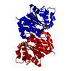
| ||||||||||||
| Unit cell |
|
-Components
| #1: Protein | Mass: 20271.391 Da / Num. of mol.: 1 Source method: isolated from a genetically manipulated source Source: (gene. exp.) Homo sapiens (human) / Gene: PARK7 / Plasmid: pET15b / Production host:  References: UniProt: Q99497, Hydrolases; Acting on ester bonds; Thioester hydrolases, Hydrolases; Acting on carbon-nitrogen bonds, other than peptide bonds; In linear amides, protein deglycase |
|---|---|
| #2: Water | ChemComp-HOH /  Mass: 18.015 Da / Num. of mol.: 124 / Source method: isolated from a natural source / Formula: H2O Mass: 18.015 Da / Num. of mol.: 124 / Source method: isolated from a natural source / Formula: H2O |
| Has ligand of interest | Y |
| Has protein modification | Y |
-Experimental details
-Experiment
| Experiment | Method: X-RAY DIFFRACTION / Number of used crystals: 1 |
|---|
- Sample preparation
| Crystal | Density Matthews: 3.15 Å3/Da / Density % sol: 60.89 % / Description: 20-25 micron microcrystals |
|---|---|
| Crystal grow | Temperature: 293 K / Method: batch mode / pH: 7.5 / Details: 100 mM HEPES, 200 mM NaCl, 15% (w/v) PEG 3350 |
-Data collection
| Diffraction | Mean temperature: 293 K / Serial crystal experiment: Y | |||||||||
|---|---|---|---|---|---|---|---|---|---|---|
| Diffraction source | Source: SYNCHROTRON / Site: APS / Beamline: 14-ID-B / Wavelength: 1.240-1.016 | |||||||||
| Detector | Type: RAYONIX MX340-HS / Detector: CCD / Date: Nov 13, 2022 | |||||||||
| Radiation | Protocol: LAUE / Monochromatic (M) / Laue (L): L / Scattering type: x-ray | |||||||||
| Radiation wavelength |
| |||||||||
| Reflection | Resolution: 1.77→65.84 Å / Num. obs: 24998 / % possible obs: 99.01 % / Redundancy: 95.32 % / Biso Wilson estimate: 27.55 Å2 / CC1/2: 0.99 / R split: 0.101 / Net I/σ(I): 4.68 | |||||||||
| Reflection shell | Resolution: 1.77→1.8 Å / Redundancy: 90.5 % / Mean I/σ(I) obs: 0.31 / Num. unique obs: 2428 / CC1/2: 0.05 / % possible all: 98.7 | |||||||||
| Serial crystallography sample delivery | Description: concentric flow microfluidic mixer / Method: injection | |||||||||
| Serial crystallography data reduction | Crystal hits: 1518 / Frames indexed: 1477 / Frames total: 20000 |
- Processing
| Software |
| |||||||||||||||||||||||||||||||||||||||||||||||||
|---|---|---|---|---|---|---|---|---|---|---|---|---|---|---|---|---|---|---|---|---|---|---|---|---|---|---|---|---|---|---|---|---|---|---|---|---|---|---|---|---|---|---|---|---|---|---|---|---|---|---|
| Refinement | Method to determine structure: FOURIER SYNTHESIS / Resolution: 1.97→32.96 Å / SU ML: 0.1935 / Cross valid method: FREE R-VALUE / σ(F): 1.34 / Phase error: 20.9654 Stereochemistry target values: GeoStd + Monomer Library + CDL v1.2
| |||||||||||||||||||||||||||||||||||||||||||||||||
| Solvent computation | Shrinkage radii: 0.9 Å / VDW probe radii: 1.11 Å / Solvent model: FLAT BULK SOLVENT MODEL | |||||||||||||||||||||||||||||||||||||||||||||||||
| Displacement parameters | Biso mean: 34.46 Å2 | |||||||||||||||||||||||||||||||||||||||||||||||||
| Refinement step | Cycle: LAST / Resolution: 1.97→32.96 Å
| |||||||||||||||||||||||||||||||||||||||||||||||||
| Refine LS restraints |
| |||||||||||||||||||||||||||||||||||||||||||||||||
| LS refinement shell |
|
