Movie
Movie Controller


+ Open data

- Basic information
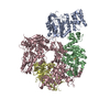
| Entry | Database: PDB / ID: 9c8v | ||||||||||||
|---|---|---|---|---|---|---|---|---|---|---|---|---|---|
| Title | Human DNA polymerase alpha/primase - CHAPSO (4 mM) | ||||||||||||
 Components Components |
| ||||||||||||
 Keywords Keywords | REPLICATION / Human DNA poly alpha/primase / CHAPSO | ||||||||||||
| Function / homology |  Function and homology information Function and homology informationribonucleotide binding / DNA primase AEP / DNA replication initiation / DNA/RNA hybrid binding / Inhibition of replication initiation of damaged DNA by RB1/E2F1 / alpha DNA polymerase:primase complex / regulation of type I interferon production / Telomere C-strand synthesis initiation / Polymerase switching / Processive synthesis on the lagging strand ...ribonucleotide binding / DNA primase AEP / DNA replication initiation / DNA/RNA hybrid binding / Inhibition of replication initiation of damaged DNA by RB1/E2F1 / alpha DNA polymerase:primase complex / regulation of type I interferon production / Telomere C-strand synthesis initiation / Polymerase switching / Processive synthesis on the lagging strand / Removal of the Flap Intermediate / lagging strand elongation / DNA replication, synthesis of primer / mitotic DNA replication initiation / Polymerase switching on the C-strand of the telomere / DNA strand elongation involved in DNA replication / leading strand elongation / G1/S-Specific Transcription / DNA synthesis involved in DNA repair / DNA replication origin binding / Activation of the pre-replicative complex / DNA replication initiation / Defective pyroptosis / double-strand break repair via nonhomologous end joining / nuclear matrix / protein import into nucleus / DNA-directed RNA polymerase activity / nuclear envelope / single-stranded DNA binding / 4 iron, 4 sulfur cluster binding / DNA-directed DNA polymerase / DNA-directed DNA polymerase activity / DNA replication / ciliary basal body / nucleotide binding / DNA repair / chromatin binding / protein kinase binding / chromatin / nucleolus / magnesium ion binding / DNA binding / zinc ion binding / nucleoplasm / membrane / metal ion binding / nucleus / cytosol Similarity search - Function | ||||||||||||
| Biological species |  Homo sapiens (human) Homo sapiens (human) | ||||||||||||
| Method | ELECTRON MICROSCOPY / single particle reconstruction / cryo EM / Resolution: 3.39 Å | ||||||||||||
 Authors Authors | Abe, K.M. / Li, G. / He, Q. / Grant, T. / Lim, C. | ||||||||||||
| Funding support |  United States, 3items United States, 3items
| ||||||||||||
 Citation Citation | Journal: Nat Commun / Year: 2024 Title: Small LEA proteins mitigate air-water interface damage to fragile cryo-EM samples during plunge freezing. Authors: Kaitlyn M Abe / Gan Li / Qixiang He / Timothy Grant / Ci Ji Lim / Abstract: Air-water interface (AWI) interactions during cryo-electron microscopy (cryo-EM) sample preparation cause significant sample loss, hindering structural biology research. Organisms like nematodes and ...Air-water interface (AWI) interactions during cryo-electron microscopy (cryo-EM) sample preparation cause significant sample loss, hindering structural biology research. Organisms like nematodes and tardigrades produce Late Embryogenesis Abundant (LEA) proteins to withstand desiccation stress. Here we show that these LEA proteins, when used as additives during plunge freezing, effectively mitigate AWI damage to fragile multi-subunit molecular samples. The resulting high-resolution cryo-EM maps are comparable to or better than those obtained using existing AWI damage mitigation methods. Cryogenic electron tomography reveals that particles are localized at specific interfaces, suggesting LEA proteins form a barrier at the AWI. This interaction may explain the observed sample-dependent preferred orientation of particles. LEA proteins offer a simple, cost-effective, and adaptable approach for cryo-EM structural biologists to overcome AWI-related sample damage, potentially revitalizing challenging projects and advancing the field of structural biology. | ||||||||||||
| History |
|
- Structure visualization
| Structure viewer | Molecule: MolmilJmol/JSmol |
|---|
- Downloads & links
-Download
| PDBx/mmCIF format | 9c8v.cif.gz | 585.1 KB | Display | PDBx/mmCIF format |
|---|---|---|---|---|
| PDB format | pdb9c8v.ent.gz | 376 KB | Display | PDB format |
| PDBx/mmJSON format | 9c8v.json.gz | Tree view | PDBx/mmJSON format | |
| Others |  Other downloads Other downloads |
-Validation report
| Arichive directory | https://data.pdbj.org/pub/pdb/validation_reports/c8/9c8vftp://data.pdbj.org/pub/pdb/validation_reports/c8/9c8v | HTTPS FTP |
|---|
-Related structure data
| Related structure data |  43627MC  8vy3C 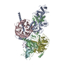 9c8uC C: citing same article ( M: map data used to model this data |
|---|---|
| Similar structure data |


-Links
 PDBj
PDBj


- Assembly
| Deposited unit | 
|
|---|---|
| 1 |
|
-Components
-Protein , 2 types, 2 molecules AB
| #1: Protein | Mass: 49016.941 Da / Num. of mol.: 1 Source method: isolated from a genetically manipulated source Source: (gene. exp.) Homo sapiens (human) / Gene: PRIM1 / Production host:  Trichoplusia ni (cabbage looper) / References: UniProt: P49642, DNA primase AEP Trichoplusia ni (cabbage looper) / References: UniProt: P49642, DNA primase AEP |
|---|---|
| #2: Protein | Mass: 50538.652 Da / Num. of mol.: 1 Source method: isolated from a genetically manipulated source Source: (gene. exp.) Homo sapiens (human) / Gene: PRIM2, PRIM2A / Production host: Trichoplusia ni (cabbage looper) / References: UniProt: P49643 |
-DNA polymerase alpha ... , 2 types, 2 molecules CD
| #3: Protein | Mass: 128446.852 Da / Num. of mol.: 1 Source method: isolated from a genetically manipulated source Source: (gene. exp.) Homo sapiens (human) / Gene: POLA1, POLA / Production host: Trichoplusia ni (cabbage looper) / References: UniProt: P09884, DNA-directed DNA polymerase |
|---|---|
| #4: Protein | Mass: 49074.590 Da / Num. of mol.: 1 Source method: isolated from a genetically manipulated source Source: (gene. exp.) Homo sapiens (human) / Gene: POLA2 / Production host: Trichoplusia ni (cabbage looper) / References: UniProt: Q14181 |
-Non-polymers , 2 types, 4 molecules 

| #5: Chemical |  Mass: 65.409 Da / Num. of mol.: 3 / Source method: obtained synthetically / Formula: Zn Mass: 65.409 Da / Num. of mol.: 3 / Source method: obtained synthetically / Formula: Zn#6: Chemical | ChemComp-SF4 / |  Mass: 351.640 Da / Num. of mol.: 1 / Source method: obtained synthetically / Formula: Fe4S4 Mass: 351.640 Da / Num. of mol.: 1 / Source method: obtained synthetically / Formula: Fe4S4 |
|---|
-Details
| Has ligand of interest | N |
|---|---|
| Has protein modification | N |
-Experimental details
-Experiment
| Experiment | Method: ELECTRON MICROSCOPY |
|---|---|
| EM experiment | Aggregation state: PARTICLE / 3D reconstruction method: single particle reconstruction |
- Sample preparation
| Component | Name: Human DNA polymerase alpha/primase - CHAPSO (4 mM) / Type: COMPLEX / Entity ID: #1-#4 / Source: RECOMBINANT |
|---|---|
| Source (natural) | Organism: Homo sapiens (human) |
| Source (recombinant) | Organism: Trichoplusia ni (cabbage looper) |
| Buffer solution | pH: 7 |
| Specimen | Conc.: 4.69 mg/ml / Embedding applied: NO / Shadowing applied: NO / Staining applied: NO / Vitrification applied: YES |
| Vitrification | Cryogen name: ETHANE / Humidity: 95 % |
- Electron microscopy imaging
| Experimental equipment |  Model: Talos Arctica / Image courtesy: FEI Company |
|---|---|
| Microscopy | Model: FEI TALOS ARCTICA |
| Electron gun | Electron source:  FIELD EMISSION GUN / Accelerating voltage: 200 kV / Illumination mode: FLOOD BEAM FIELD EMISSION GUN / Accelerating voltage: 200 kV / Illumination mode: FLOOD BEAM |
| Electron lens | Mode: BRIGHT FIELD / Nominal defocus max: 2500 nm / Nominal defocus min: 1000 nm |
| Image recording | Electron dose: 50 e/Å2 / Film or detector model: GATAN K3 BIOQUANTUM (6k x 4k) |
- Processing
| EM software | Name: PHENIX / Version: 1.21.1_5286 / Category: model refinement | ||||||||||||||||||||||||
|---|---|---|---|---|---|---|---|---|---|---|---|---|---|---|---|---|---|---|---|---|---|---|---|---|---|
| CTF correction | Type: NONE | ||||||||||||||||||||||||
| 3D reconstruction | Resolution: 3.39 Å / Resolution method: FSC 0.143 CUT-OFF / Num. of particles: 674789 / Symmetry type: POINT | ||||||||||||||||||||||||
| Atomic model building | PDB-ID: 5exr Accession code: 5exr / Source name: PDB / Type: experimental model | ||||||||||||||||||||||||
| Refinement | Cross valid method: NONE Stereochemistry target values: GeoStd + Monomer Library + CDL v1.2 | ||||||||||||||||||||||||
| Displacement parameters | Biso mean: 17.95 Å2 | ||||||||||||||||||||||||
| Refine LS restraints |
|