 Movie
Movie Controller


+ Open data

- Basic information
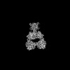
| Entry | Database: PDB / ID: 9c60 | |||||||||||||||||||||
|---|---|---|---|---|---|---|---|---|---|---|---|---|---|---|---|---|---|---|---|---|---|---|
| Title | CryoEM structure of kainate receptor Gluk2 in apo state | |||||||||||||||||||||
 Components Components | Glutamate receptor ionotropic, kainate 2 | |||||||||||||||||||||
 Keywords Keywords | STRUCTURAL PROTEIN / C1 / membrane / homotetrmer / glutamate | |||||||||||||||||||||
| Function / homology |  Function and homology information Function and homology informationmossy fiber rosette / detection of cold stimulus involved in thermoception / Activation of Na-permeable kainate receptors / Activation of Ca-permeable Kainate Receptor / kainate selective glutamate receptor complex / regulation of short-term neuronal synaptic plasticity / ubiquitin conjugating enzyme binding / glutamate receptor activity / negative regulation of synaptic transmission, glutamatergic / regulation of JNK cascade ...mossy fiber rosette / detection of cold stimulus involved in thermoception / Activation of Na-permeable kainate receptors / Activation of Ca-permeable Kainate Receptor / kainate selective glutamate receptor complex / regulation of short-term neuronal synaptic plasticity / ubiquitin conjugating enzyme binding / glutamate receptor activity / negative regulation of synaptic transmission, glutamatergic / regulation of JNK cascade / receptor clustering / inhibitory postsynaptic potential / kainate selective glutamate receptor activity / extracellularly glutamate-gated ion channel activity / modulation of excitatory postsynaptic potential / ionotropic glutamate receptor complex / positive regulation of synaptic transmission / behavioral fear response / neuronal action potential / glutamate-gated receptor activity / glutamate-gated calcium ion channel activity / dendrite cytoplasm / ligand-gated monoatomic ion channel activity involved in regulation of presynaptic membrane potential / presynaptic modulation of chemical synaptic transmission / hippocampal mossy fiber to CA3 synapse / SNARE binding / PDZ domain binding / transmitter-gated monoatomic ion channel activity involved in regulation of postsynaptic membrane potential / synaptic transmission, glutamatergic / regulation of membrane potential / excitatory postsynaptic potential / intracellular protein transport / regulation of long-term neuronal synaptic plasticity / postsynaptic density membrane / modulation of chemical synaptic transmission / intracellular calcium ion homeostasis / terminal bouton / positive regulation of neuron apoptotic process / neuron apoptotic process / scaffold protein binding / presynaptic membrane / chemical synaptic transmission / negative regulation of neuron apoptotic process / perikaryon / postsynaptic membrane / postsynaptic density / axon / neuronal cell body / ubiquitin protein ligase binding / synapse / dendrite / glutamatergic synapse / membrane / identical protein binding / plasma membrane Similarity search - Function | |||||||||||||||||||||
| Biological species |  | |||||||||||||||||||||
| Method | ELECTRON MICROSCOPY / single particle reconstruction / cryo EM / Resolution: 3.8 Å | |||||||||||||||||||||
 Authors Authors | Changping, Z. / Nami, T. | |||||||||||||||||||||
| Funding support |  United States, 2items United States, 2items
| |||||||||||||||||||||
 Citation Citation | Journal: Nat Struct Mol Biol / Year: 2025 Title: Structural basis of GluK2 kainate receptor activation by a partial agonist. Authors: Guadalupe Segura-Covarrubias / Changping Zhou / Nebojša Bogdanović / Lisa Zhang / Nami Tajima / Abstract: Kainate receptors (KARs) belong to the family of ionotropic glutamate receptors that regulate neurotransmitter release and excitatory synaptic transmission in the central nervous system. Despite ...Kainate receptors (KARs) belong to the family of ionotropic glutamate receptors that regulate neurotransmitter release and excitatory synaptic transmission in the central nervous system. Despite their critical roles in synaptic signaling and disease, the detailed gating mechanisms of KARs are not completely understood. Here we present cryo-electron microscopy structures of homomeric rat GluK2 KAR in an unliganded apo state and in complexes with a partial agonist, domoate. Partial agonist-bound GluK2 populates multiple conformations, including intermediate and desensitized states. Moreover, we demonstrate that the N-glycans at the amino-terminal domain-ligand binding domain (LBD) interface modulate receptor gating properties by interfering with cation binding at the LBD dimer interface. Together, these results provide insights into the unique gating mechanisms of KARs. | |||||||||||||||||||||
| History |
|
- Structure visualization
| Structure viewer | Molecule: MolmilJmol/JSmol |
|---|
- Downloads & links
-Download
| PDBx/mmCIF format | 9c60.cif.gz | 525.9 KB | Display | PDBx/mmCIF format |
|---|---|---|---|---|
| PDB format | pdb9c60.ent.gz | 427.4 KB | Display | PDB format |
| PDBx/mmJSON format | 9c60.json.gz | Tree view | PDBx/mmJSON format | |
| Others |  Other downloads Other downloads |
-Validation report
| Arichive directory | https://data.pdbj.org/pub/pdb/validation_reports/c6/9c60ftp://data.pdbj.org/pub/pdb/validation_reports/c6/9c60 | HTTPS FTP |
|---|
-Related structure data
| Related structure data |  45239MC  8gc5C  9c5yC  9c5zC  9cazC M: map data used to model this data C: citing same article ( |
|---|---|
| Similar structure data |


-Links
 PDBj
PDBj

- Assembly
| Deposited unit | 
|
|---|---|
| 1 |
|
-Components
| #1: Protein | Mass: 102584.031 Da / Num. of mol.: 4 Source method: isolated from a genetically manipulated source Source: (gene. exp.)  Homo sapiens (human) / References: UniProt: P42260 Homo sapiens (human) / References: UniProt: P42260#2: Sugar | ChemComp-NAG /   Type: D-saccharide, beta linking / Mass: 221.208 Da / Num. of mol.: 12 / Source method: obtained synthetically / Formula: C8H15NO6 / Feature type: SUBJECT OF INVESTIGATION Type: D-saccharide, beta linking / Mass: 221.208 Da / Num. of mol.: 12 / Source method: obtained synthetically / Formula: C8H15NO6 / Feature type: SUBJECT OF INVESTIGATIONHas ligand of interest | Y | Has protein modification | Y | |
|---|
-Experimental details
-Experiment
| Experiment | Method: ELECTRON MICROSCOPY |
|---|---|
| EM experiment | Aggregation state: PARTICLE / 3D reconstruction method: single particle reconstruction |
- Sample preparation
| Component | Name: Gluk2 homotetramer / Type: COMPLEX / Entity ID: #1 / Source: RECOMBINANT | ||||||||||||||||||||
|---|---|---|---|---|---|---|---|---|---|---|---|---|---|---|---|---|---|---|---|---|---|
| Source (natural) | Organism: | ||||||||||||||||||||
| Source (recombinant) | Organism: Homo sapiens (human) | ||||||||||||||||||||
| Buffer solution | pH: 8 | ||||||||||||||||||||
| Buffer component |
| ||||||||||||||||||||
| Specimen | Conc.: 3.4 mg/ml / Embedding applied: NO / Shadowing applied: NO / Staining applied: NO / Vitrification applied: YES | ||||||||||||||||||||
| Specimen support | Grid material: GOLD / Grid mesh size: 300 divisions/in. / Grid type: EMS Lacey Carbon | ||||||||||||||||||||
| Vitrification | Instrument: FEI VITROBOT MARK II / Cryogen name: NITROGEN / Humidity: 100 % / Chamber temperature: 277.15 K |
- Electron microscopy imaging
| Experimental equipment |  Model: Titan Krios / Image courtesy: FEI Company |
|---|---|
| Microscopy | Model: FEI TITAN KRIOS |
| Electron gun | Electron source:  FIELD EMISSION GUN / Accelerating voltage: 300 kV / Illumination mode: FLOOD BEAM FIELD EMISSION GUN / Accelerating voltage: 300 kV / Illumination mode: FLOOD BEAM |
| Electron lens | Mode: BRIGHT FIELD / Nominal magnification: 81000 X / Calibrated magnification: 81000 X / Nominal defocus max: 1600 nm / Nominal defocus min: 800 nm / Calibrated defocus min: 800 nm / Calibrated defocus max: 1600 nm / Cs: 2.7 mm / C2 aperture diameter: 51 µm |
| Image recording | Average exposure time: 1 sec. / Electron dose: 50 e/Å2 / Film or detector model: GATAN K3 BIOQUANTUM (6k x 4k) |
- Processing
| EM software |
| ||||||||||||||||||||||||
|---|---|---|---|---|---|---|---|---|---|---|---|---|---|---|---|---|---|---|---|---|---|---|---|---|---|
| CTF correction | Type: PHASE FLIPPING AND AMPLITUDE CORRECTION | ||||||||||||||||||||||||
| Particle selection | Num. of particles selected: 1198495 | ||||||||||||||||||||||||
| Symmetry | Point symmetry: C1 (asymmetric) | ||||||||||||||||||||||||
| 3D reconstruction | Resolution: 3.8 Å / Resolution method: FSC 0.143 CUT-OFF / Num. of particles: 171688 / Algorithm: FOURIER SPACE / Symmetry type: POINT | ||||||||||||||||||||||||
| Atomic model building | Protocol: RIGID BODY FIT | ||||||||||||||||||||||||
| Refine LS restraints |
|
