 Movie
Movie Controller

[English] 日本語
 Yorodumi
- PDB-9c52: Cryo-EM structure of the Strand displacement Complex (I) of Yeast... -
+ Open data

- Basic information
| Entry | Database: PDB / ID: 9c52 | ||||||
|---|---|---|---|---|---|---|---|
| Title | Cryo-EM structure of the Strand displacement Complex (I) of Yeast Mitochondrial DNA polymerase Gamma (MIP1) with downstream DNA | ||||||
 Components Components |
| ||||||
 Keywords Keywords | Transferase/DNA / Mitochondrial DNA Polymerase Gamma / Strand displacement complex / MIP1 / REPLICATION / Helicase independent DNA polymerase / Transferase-DNA complex | ||||||
| Function / homology | 3'-DEOXYTHYMIDINE-5'-MONOPHOSPHATE / 2'-DEOXYADENOSINE 5'-TRIPHOSPHATE / DNA / DNA (> 10) / :  Function and homology information Function and homology information | ||||||
| Biological species |  synthetic construct (others) | ||||||
| Method | ELECTRON MICROSCOPY / single particle reconstruction / cryo EM / Resolution: 2.64 Å | ||||||
 Authors Authors | Nayak, A.R. / Sokolova, V.O. / Sillamaa, S. / Sedmen, J. / Temiakov, D. | ||||||
| Funding support |  United States, 1items United States, 1items
| ||||||
 Citation Citation | Journal: Nat Commun / Year: 2025 Title: Structural basis for intrinsic strand displacement activity of mitochondrial DNA polymerase. Authors: Ashok R Nayak / Viktoriia Sokolova / Sirelin Sillamaa / Karl Herbine / Juhan Sedman / Dmitry Temiakov /  Abstract: Members of the Pol A family of DNA polymerases, found across all domains of life, utilize various strategies for DNA strand separation during replication. In higher eukaryotes, mitochondrial DNA ...Members of the Pol A family of DNA polymerases, found across all domains of life, utilize various strategies for DNA strand separation during replication. In higher eukaryotes, mitochondrial DNA polymerase γ relies on the replicative helicase TWINKLE, whereas the yeast ortholog, Mip1, can unwind DNA independently. Using Mip1 as a model, we present a series of high-resolution cryo-EM structures that capture the process of DNA strand displacement. Our data reveal previously unidentified structural elements that facilitate the unwinding of the downstream DNA duplex. Yeast cells harboring Mip1 variants defective in strand displacement exhibit impaired oxidative phosphorylation and loss of mtDNA, corroborating the structural observations. This study provides a molecular basis for the intrinsic strand displacement activity of Mip1 and illuminates the distinct unwinding mechanisms utilized by Pol A family DNA polymerases. | ||||||
| History |
|
- Structure visualization
| Structure viewer | Molecule: MolmilJmol/JSmol |
|---|
- Downloads & links
-Download
| PDBx/mmCIF format | 9c52.cif.gz | 228.2 KB | Display | PDBx/mmCIF format |
|---|---|---|---|---|
| PDB format | pdb9c52.ent.gz | 167.8 KB | Display | PDB format |
| PDBx/mmJSON format | 9c52.json.gz | Tree view | PDBx/mmJSON format | |
| Others |  Other downloads Other downloads |
-Validation report
| Arichive directory | https://data.pdbj.org/pub/pdb/validation_reports/c5/9c52ftp://data.pdbj.org/pub/pdb/validation_reports/c5/9c52 | HTTPS FTP |
|---|
-Related structure data
| Related structure data | 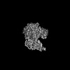 45195MC  9c51C  9c53C 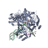 9d2iC M: map data used to model this data C: citing same article ( |
|---|---|
| Similar structure data |


-Links
 PDBj
PDBj


- Assembly
| Deposited unit | 
|
|---|---|
| 1 |
|
-Components
-Protein , 1 types, 1 molecules A
| #1: Protein | Mass: 141745.438 Da / Num. of mol.: 1 Source method: isolated from a genetically manipulated source Source: (gene. exp.) Gene: MIP1, GI527_G0005701 / Production host:  References: UniProt: A0A8H4BW69, DNA-directed DNA polymerase |
|---|
-DNA chain , 3 types, 3 molecules NTP
| #2: DNA chain | Mass: 7078.555 Da / Num. of mol.: 1 / Source method: obtained synthetically / Source: (synth.) synthetic construct (others) |
|---|---|
| #3: DNA chain | Mass: 12854.218 Da / Num. of mol.: 1 / Source method: obtained synthetically / Source: (synth.) synthetic construct (others) |
| #4: DNA chain | Mass: 7172.610 Da / Num. of mol.: 1 / Source method: obtained synthetically / Source: (synth.) synthetic construct (others) |
-Non-polymers , 3 types, 3 molecules 

| #5: Chemical | ChemComp-MG /  Mass: 24.305 Da / Num. of mol.: 1 / Source method: obtained synthetically / Formula: Mg Mass: 24.305 Da / Num. of mol.: 1 / Source method: obtained synthetically / Formula: Mg |
|---|---|
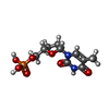
| #6: Chemical | ChemComp-DTP /  Mass: 491.182 Da / Num. of mol.: 1 / Source method: obtained synthetically / Formula: C10H16N5O12P3 / Feature type: SUBJECT OF INVESTIGATION Mass: 491.182 Da / Num. of mol.: 1 / Source method: obtained synthetically / Formula: C10H16N5O12P3 / Feature type: SUBJECT OF INVESTIGATION |
| #7: Chemical | ChemComp-2DT /  Type: DNA linking / Mass: 306.209 Da / Num. of mol.: 1 / Source method: obtained synthetically / Formula: C10H15N2O7P / Feature type: SUBJECT OF INVESTIGATION Type: DNA linking / Mass: 306.209 Da / Num. of mol.: 1 / Source method: obtained synthetically / Formula: C10H15N2O7P / Feature type: SUBJECT OF INVESTIGATION |
-Details
| Has ligand of interest | Y |
|---|---|
| Has protein modification | N |
-Experimental details
-Experiment
| Experiment | Method: ELECTRON MICROSCOPY |
|---|---|
| EM experiment | Aggregation state: PARTICLE / 3D reconstruction method: single particle reconstruction |
- Sample preparation
| Component | Name: Cryo-EM map of the Strand displacement Complex (II) of Yeast Mitochondrial DNA polymerase Gamma Type: COMPLEX Details: Strand displacement complex of yeast Mitochondrial DNA polymerase Gamma (MIP1) wild type assembled on a primer-template and a downstream non-template strand. Entity ID: #1-#4 / Source: RECOMBINANT |
|---|---|
| Molecular weight | Value: 0.142 MDa / Experimental value: YES |
| Source (natural) | Organism: |
| Source (recombinant) | Organism: |
| Buffer solution | pH: 7.9 / Details: 10 mM Tris-HCL, 100 mM NaCL, 10 mM DTT, 5 mM MgCl2 |
| Specimen | Conc.: 0.57 mg/ml / Embedding applied: NO / Shadowing applied: NO / Staining applied: NO / Vitrification applied: YES Details: A 4 uM strand displacement complex was assembled with wild-type Mip1 and a DNA scaffold with primer, template, and non-template strands, mixed in equal millimolar ratio. The primer strand ...Details: A 4 uM strand displacement complex was assembled with wild-type Mip1 and a DNA scaffold with primer, template, and non-template strands, mixed in equal millimolar ratio. The primer strand was extended by three nucleotides with 1 mM dGTP and chain terminated with 1 mM di-deoxy TTP. 1 mM dATP was added before plunge freezing. |
| Specimen support | Grid material: COPPER / Grid mesh size: 300 divisions/in. / Grid type: Quantifoil R1.2/1.3 |
| Vitrification | Instrument: FEI VITROBOT MARK IV / Cryogen name: ETHANE / Humidity: 95 % / Chamber temperature: 277 K |
- Electron microscopy imaging
| Experimental equipment |  Model: Titan Krios / Image courtesy: FEI Company |
|---|---|
| Microscopy | Model: FEI TITAN KRIOS |
| Electron gun | Electron source:  FIELD EMISSION GUN / Accelerating voltage: 300 kV / Illumination mode: FLOOD BEAM FIELD EMISSION GUN / Accelerating voltage: 300 kV / Illumination mode: FLOOD BEAM |
| Electron lens | Mode: BRIGHT FIELD / Nominal magnification: 105000 X / Nominal defocus max: 1500 nm / Nominal defocus min: 500 nm / Cs: 2.7 mm |
| Specimen holder | Cryogen: NITROGEN / Specimen holder model: FEI TITAN KRIOS AUTOGRID HOLDER |
| Image recording | Electron dose: 60 e/Å2 / Film or detector model: FEI FALCON IV (4k x 4k) / Num. of grids imaged: 2 / Num. of real images: 14819 |
| EM imaging optics | Energyfilter slit width: 10 eV |
- Processing
| EM software |
| ||||||||||||||||||||||||||||||||
|---|---|---|---|---|---|---|---|---|---|---|---|---|---|---|---|---|---|---|---|---|---|---|---|---|---|---|---|---|---|---|---|---|---|
| Image processing | Details: Falcon 4i | ||||||||||||||||||||||||||||||||
| CTF correction | Details: Patch CTF estimation in cryoSPARC. / Type: PHASE FLIPPING AND AMPLITUDE CORRECTION | ||||||||||||||||||||||||||||||||
| Particle selection | Num. of particles selected: 9600000 | ||||||||||||||||||||||||||||||||
| Symmetry | Point symmetry: C1 (asymmetric) | ||||||||||||||||||||||||||||||||
| 3D reconstruction | Resolution: 2.64 Å / Resolution method: FSC 0.143 CUT-OFF / Num. of particles: 346877 / Num. of class averages: 1 / Symmetry type: POINT | ||||||||||||||||||||||||||||||||
| Atomic model building | Protocol: FLEXIBLE FIT / Space: REAL | ||||||||||||||||||||||||||||||||
| Atomic model building | Accession code: AF-P15801-F1 / Chain residue range: 1-1254 / Source name: AlphaFold / Type: in silico model | ||||||||||||||||||||||||||||||||
| Refine LS restraints |
|
