Movie
Movie Controller


+ Open data

- Basic information
| Entry | Database: PDB / ID: 9c4h | ||||||
|---|---|---|---|---|---|---|---|
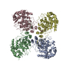
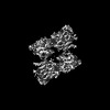
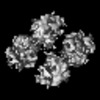
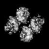
| Title | Double helical structure of influenza D RNP complex | ||||||
 Components Components |
| ||||||
 Keywords Keywords | VIRAL PROTEIN / RNA BINDING PROTEIN/RNA / Influenza / Ribonucleoprotein complex / nucleoprotein / RNA BINDING PROTEIN-RNA complex | ||||||
| Function / homology |  Function and homology information Function and homology informationhelical viral capsid / viral penetration into host nucleus / host cell / viral nucleocapsid / ribonucleoprotein complex / symbiont entry into host cell / structural molecule activity / RNA binding Similarity search - Function | ||||||
| Biological species |  Influenza D virus Influenza D virus | ||||||
| Method | ELECTRON MICROSCOPY / helical reconstruction / cryo EM / Resolution: 8.6 Å | ||||||
 Authors Authors | Peng, R. / Chang, Y.-W. | ||||||
| Funding support |  United States, 1items United States, 1items
| ||||||
 Citation Citation | Journal: Science / Year: 2025 Title: Molecular basis of influenza ribonucleoprotein complex assembly and processive RNA synthesis. Authors: Ruchao Peng / Xin Xu / Binod Nepal / Yikang Gong / Fenglin Li / Max B Ferretti / Mingyang Zhou / Kristen W Lynch / George M Burslem / Sandhya Kortagere / Ronen Marmorstein / Yi-Wei Chang / Abstract: Influenza viruses replicate and transcribe their genome in the context of a conserved ribonucleoprotein (RNP) complex. By integrating cryo-electron microscopy single-particle analysis and cryo- ...Influenza viruses replicate and transcribe their genome in the context of a conserved ribonucleoprotein (RNP) complex. By integrating cryo-electron microscopy single-particle analysis and cryo-electron tomography, we define the influenza RNP as a right-handed, antiparallel double helix with the viral RNA encapsidated in the minor groove. Individual nucleoprotein subunits are connected by a flexible tail loop that inserts into a conserved pocket in its neighbor. We visualize the viral polymerase in RNP at different functional states, revealing how it accesses the RNA template while maintaining the double-helical architecture of RNP by strand sliding. Targeting the tail loop binding interface, we identify lead compounds as potential anti-influenza inhibitors. These findings elucidate the molecular determinants underpinning influenza virus replication and highlight a promising target for antiviral development. | ||||||
| History |
|
- Structure visualization
| Structure viewer | Molecule: MolmilJmol/JSmol |
|---|
- Downloads & links
-Download
| PDBx/mmCIF format | 9c4h.cif.gz | 1.4 MB | Display | PDBx/mmCIF format |
|---|---|---|---|---|
| PDB format | pdb9c4h.ent.gz | 1.2 MB | Display | PDB format |
| PDBx/mmJSON format | 9c4h.json.gz | Tree view | PDBx/mmJSON format | |
| Others |  Other downloads Other downloads |
-Validation report
| Arichive directory | https://data.pdbj.org/pub/pdb/validation_reports/c4/9c4hftp://data.pdbj.org/pub/pdb/validation_reports/c4/9c4h | HTTPS FTP |
|---|
-Related structure data
| Related structure data |  44980MC  9bwvC  9bwzC  9bx0C  9bx1C  9bx4C M: map data used to model this data C: citing same article ( |
|---|---|
| Similar structure data |


-Links
 PDBj
PDBj


- Assembly
| Deposited unit | 
|
|---|---|
| 1 |
|
-Components
| #1: Protein | Mass: 61329.227 Da / Num. of mol.: 16 Source method: isolated from a genetically manipulated source Source: (gene. exp.) Influenza D virus / Gene: NP / Production host:  Homo sapiens (human) / References: UniProt: K9LG94 Homo sapiens (human) / References: UniProt: K9LG94#2: RNA chain | | Mass: 265709.594 Da / Num. of mol.: 1 Source method: isolated from a genetically manipulated source Source: (gene. exp.) Influenza D virus / Production host: Homo sapiens (human)Has protein modification | N | |
|---|
-Experimental details
-Experiment
| Experiment | Method: ELECTRON MICROSCOPY |
|---|---|
| EM experiment | Aggregation state: PARTICLE / 3D reconstruction method: helical reconstruction |
- Sample preparation
| Component | Name: Influenza D virus RNP complex / Type: COMPLEX / Entity ID: all / Source: RECOMBINANT |
|---|---|
| Molecular weight | Experimental value: NO |
| Source (natural) | Organism: Influenza D virus |
| Source (recombinant) | Organism: Homo sapiens (human) |
| Buffer solution | pH: 7.4 / Details: 1x PBS |
| Specimen | Embedding applied: NO / Shadowing applied: NO / Staining applied: NO / Vitrification applied: YES |
| Specimen support | Grid material: GOLD / Grid mesh size: 300 divisions/in. / Grid type: Quantifoil R1.2/1.3 |
| Vitrification | Instrument: LEICA EM GP / Cryogen name: ETHANE-PROPANE |
- Electron microscopy imaging
| Experimental equipment |  Model: Titan Krios / Image courtesy: FEI Company |
|---|---|
| Microscopy | Model: TFS KRIOS |
| Electron gun | Electron source:  FIELD EMISSION GUN / Accelerating voltage: 300 kV / Illumination mode: FLOOD BEAM FIELD EMISSION GUN / Accelerating voltage: 300 kV / Illumination mode: FLOOD BEAM |
| Electron lens | Mode: BRIGHT FIELD / Nominal defocus max: 3500 nm / Nominal defocus min: 1300 nm / Cs: 2.7 mm / C2 aperture diameter: 100 µm / Alignment procedure: COMA FREE |
| Specimen holder | Cryogen: NITROGEN / Specimen holder model: FEI TITAN KRIOS AUTOGRID HOLDER |
| Image recording | Electron dose: 60 e/Å2 / Film or detector model: GATAN K3 BIOQUANTUM (6k x 4k) |
| EM imaging optics | Energyfilter name: GIF Bioquantum / Energyfilter slit width: 20 eV |
- Processing
| EM software |
| ||||||||||||||||
|---|---|---|---|---|---|---|---|---|---|---|---|---|---|---|---|---|---|
| CTF correction | Type: PHASE FLIPPING AND AMPLITUDE CORRECTION | ||||||||||||||||
| Helical symmerty | Angular rotation/subunit: 61.64 ° / Axial rise/subunit: 26.87 Å / Axial symmetry: D1 | ||||||||||||||||
| 3D reconstruction | Resolution: 8.6 Å / Resolution method: FSC 0.143 CUT-OFF / Num. of particles: 44679 / Algorithm: FOURIER SPACE / Symmetry type: HELICAL | ||||||||||||||||
| Atomic model building | Protocol: FLEXIBLE FIT / Space: REAL / Target criteria: cross-correlation coefficient | ||||||||||||||||
| Atomic model building | PDB-ID: 5N2U Accession code: 5N2U / Source name: PDB / Type: experimental model |