 Movie
Movie Controller


+ Open data

- Basic information
| Entry | Database: PDB / ID: 9c44 | ||||||
|---|---|---|---|---|---|---|---|
| Title | SARS-CoV-2 S + S2L20 | ||||||
 Components Components |
| ||||||
 Keywords Keywords | VIRAL PROTEIN / S2L20 / S / NTD / RBD / coronavirus / COVID-19 / SARS-CoV-2 / hexapro / Structural Genomics / Seattle Structural Genomics Center for Infectious Disease / SSGCID | ||||||
| Function / homology |  Function and homology information Function and homology informationsymbiont-mediated disruption of host tissue / Maturation of spike protein / Translation of Structural Proteins / Virion Assembly and Release / host cell surface / host extracellular region / symbiont-mediated-mediated suppression of host tetherin activity / Induction of Cell-Cell Fusion / structural constituent of virion / positive regulation of viral entry into host cell ...symbiont-mediated disruption of host tissue / Maturation of spike protein / Translation of Structural Proteins / Virion Assembly and Release / host cell surface / host extracellular region / symbiont-mediated-mediated suppression of host tetherin activity / Induction of Cell-Cell Fusion / structural constituent of virion / positive regulation of viral entry into host cell / membrane fusion / host cell endoplasmic reticulum-Golgi intermediate compartment membrane / Attachment and Entry / entry receptor-mediated virion attachment to host cell / receptor-mediated virion attachment to host cell / host cell surface receptor binding / symbiont-mediated suppression of host innate immune response / endocytosis involved in viral entry into host cell / receptor ligand activity / fusion of virus membrane with host plasma membrane / fusion of virus membrane with host endosome membrane / viral envelope / symbiont entry into host cell / virion attachment to host cell / host cell plasma membrane / SARS-CoV-2 activates/modulates innate and adaptive immune responses / virion membrane / membrane / identical protein binding / plasma membrane Similarity search - Function | ||||||
| Biological species |   Severe acute respiratory syndrome coronavirus 2 Severe acute respiratory syndrome coronavirus 2 Homo sapiens (human) Homo sapiens (human) | ||||||
| Method | ELECTRON MICROSCOPY / single particle reconstruction / cryo EM / Resolution: 2.6 Å | ||||||
 Authors Authors | McCallum, M. / Veesler, D. / Seattle Structural Genomics Center for Infectious Disease (SSGCID) | ||||||
| Funding support |  United States, 1items United States, 1items
| ||||||
 Citation Citation | Journal: Nature / Year: 2024 Title: Design of customized coronavirus receptors. Authors: Peng Liu / Mei-Ling Huang / Hua Guo / Matthew McCallum / Jun-Yu Si / Yuan-Mei Chen / Chun-Li Wang / Xiao Yu / Lu-Lu Shi / Qing Xiong / Cheng-Bao Ma / John E Bowen / Fei Tong / Chen Liu / Ye- ...Authors: Peng Liu / Mei-Ling Huang / Hua Guo / Matthew McCallum / Jun-Yu Si / Yuan-Mei Chen / Chun-Li Wang / Xiao Yu / Lu-Lu Shi / Qing Xiong / Cheng-Bao Ma / John E Bowen / Fei Tong / Chen Liu / Ye-Hui Sun / Xiao Yang / Jing Chen / Ming Guo / Jing Li / Davide Corti / David Veesler / Zheng-Li Shi / Huan Yan /   Abstract: Although coronaviruses use diverse receptors, the characterization of coronaviruses with unknown receptors has been impeded by a lack of infection models. Here we introduce a strategy to engineer ...Although coronaviruses use diverse receptors, the characterization of coronaviruses with unknown receptors has been impeded by a lack of infection models. Here we introduce a strategy to engineer functional customized viral receptors (CVRs). The modular design relies on building artificial receptor scaffolds comprising various modules and generating specific virus-binding domains. We identify key factors for CVRs to functionally mimic native receptors by facilitating spike proteolytic cleavage, membrane fusion, pseudovirus entry and propagation for various coronaviruses. We delineate functional SARS-CoV-2 spike receptor-binding sites for CVR design and reveal the mechanism of cell entry promoted by the N-terminal domain-targeting S2L20-CVR. We generated CVR-expressing cells for 12 representative coronaviruses from 6 subgenera, most of which lack known receptors, and show that a pan-sarbecovirus CVR supports propagation of a propagation-competent HKU3 pseudovirus and of authentic RsHuB2019A. Using an HKU5-specific CVR, we successfully rescued wild-type and ZsGreen-HiBiT-incorporated HKU5-1 (LMH03f) and isolated a HKU5 strain from bat samples. Our study demonstrates the potential of the CVR strategy for establishing native receptor-independent infection models, providing a tool for studying viruses that lack known susceptible target cells. | ||||||
| History |
|
- Structure visualization
| Structure viewer | Molecule: MolmilJmol/JSmol |
|---|
- Downloads & links
-Download
| PDBx/mmCIF format | 9c44.cif.gz | 691.9 KB | Display | PDBx/mmCIF format |
|---|---|---|---|---|
| PDB format | pdb9c44.ent.gz | 535.2 KB | Display | PDB format |
| PDBx/mmJSON format | 9c44.json.gz | Tree view | PDBx/mmJSON format | |
| Others |  Other downloads Other downloads |
-Validation report
| Arichive directory | https://data.pdbj.org/pub/pdb/validation_reports/c4/9c44ftp://data.pdbj.org/pub/pdb/validation_reports/c4/9c44 | HTTPS FTP |
|---|
-Related structure data
| Related structure data | 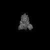 45174MC 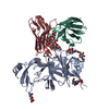 9c45C C: citing same article ( M: map data used to model this data |
|---|---|
| Similar structure data |

-Links
 PDBj
PDBj


- Assembly
| Deposited unit | 
|
|---|---|
| 1 |
|
-Components
-Protein , 1 types, 3 molecules ABC
| #1: Protein | Mass: 142427.438 Da / Num. of mol.: 3 Source method: isolated from a genetically manipulated source Source: (gene. exp.) Severe acute respiratory syndrome coronavirus 2Gene: S, 2 / Production host: Homo sapiens (human) / References: UniProt: P0DTC2 |
|---|
-Antibody , 2 types, 6 molecules HEFLDG
| #2: Antibody | Mass: 13453.958 Da / Num. of mol.: 3 Source method: isolated from a genetically manipulated source Source: (gene. exp.) Homo sapiens (human) / Gene: S, 2 / Production host: Homo sapiens (human)#3: Antibody | Mass: 11829.141 Da / Num. of mol.: 3 Source method: isolated from a genetically manipulated source Source: (gene. exp.) Homo sapiens (human) / Gene: S, 2 / Production host: Homo sapiens (human) |
|---|
-Sugars , 3 types, 45 molecules 
| #4: Polysaccharide | 2-acetamido-2-deoxy-beta-D-glucopyranose-(1-4)-2-acetamido-2-deoxy-beta-D-glucopyranose Source method: isolated from a genetically manipulated source #5: Polysaccharide | Source method: isolated from a genetically manipulated source #6: Sugar | ChemComp-NAG /  Type: D-saccharide, beta linking / Mass: 221.208 Da / Num. of mol.: 33 / Source method: obtained synthetically / Formula: C8H15NO6 Type: D-saccharide, beta linking / Mass: 221.208 Da / Num. of mol.: 33 / Source method: obtained synthetically / Formula: C8H15NO6 |
|---|
-Details
| Has ligand of interest | N |
|---|---|
| Has protein modification | Y |
-Experimental details
-Experiment
| Experiment | Method: ELECTRON MICROSCOPY |
|---|---|
| EM experiment | Aggregation state: PARTICLE / 3D reconstruction method: single particle reconstruction |
- Sample preparation
| Component | Name: SARS-CoV-2 S bound by S2L20 Fab / Type: COMPLEX / Entity ID: #1, #3, #2 / Source: RECOMBINANT |
|---|---|
| Molecular weight | Experimental value: NO |
| Source (natural) | Organism: Homo sapiens (human) |
| Source (recombinant) | Organism: Homo sapiens (human) |
| Buffer solution | pH: 8 |
| Specimen | Embedding applied: NO / Shadowing applied: NO / Staining applied: NO / Vitrification applied: YES |
| Vitrification | Cryogen name: ETHANE |
- Electron microscopy imaging
| Experimental equipment |  Model: Titan Krios / Image courtesy: FEI Company |
|---|---|
| Microscopy | Model: FEI TITAN KRIOS |
| Electron gun | Electron source:  FIELD EMISSION GUN / Accelerating voltage: 300 kV / Illumination mode: FLOOD BEAM FIELD EMISSION GUN / Accelerating voltage: 300 kV / Illumination mode: FLOOD BEAM |
| Electron lens | Mode: BRIGHT FIELD / Nominal defocus max: 20000 nm / Nominal defocus min: 2000 nm |
| Image recording | Electron dose: 63 e/Å2 / Film or detector model: GATAN K3 (6k x 4k) |
- Processing
| CTF correction | Type: PHASE FLIPPING AND AMPLITUDE CORRECTION |
|---|---|
| 3D reconstruction | Resolution: 2.6 Å / Resolution method: FSC 0.143 CUT-OFF / Num. of particles: 165733 / Symmetry type: POINT |
